 Movie
Movie Controller


+ Open data

- Basic information
| Entry | Database: PDB / ID: 3v06 | ||||||||||||||||||
|---|---|---|---|---|---|---|---|---|---|---|---|---|---|---|---|---|---|---|---|
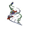
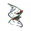
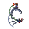
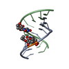
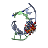
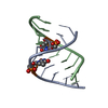
| Title | Crystal structure of S-6'-Me-3'-fluoro hexitol nucleic acid | ||||||||||||||||||
 Components Components | DNA (5'-D(* Keywords KeywordsDNA / A-form DNA / 3'-fluoro hexitol nucleic acid / FHNA / S-6'-Me-FHNA / antisense oligonucleotides | Function / homology | STRONTIUM ION / DNA |  Function and homology information Function and homology informationMethod |  X-RAY DIFFRACTION / SYNCHROTRON / MOLECULAR REPLACEMENT / Resolution: 1.53 Å X-RAY DIFFRACTION / SYNCHROTRON / MOLECULAR REPLACEMENT / Resolution: 1.53 Å  Authors AuthorsPallan, P.S. / Egli, M. |  CitationJournal: Biochemistry / Year: 2012 CitationJournal: Biochemistry / Year: 2012Title: Insights from crystal structures into the opposite effects on RNA affinity caused by the s- and R-6'-methyl backbone modifications of 3'-fluoro hexitol nucleic Acid. Authors: Pallan, P.S. / Yu, J. / Allerson, C.R. / Swayze, E.E. / Seth, P. / Egli, M. History |
|
- Structure visualization
| Structure viewer | Molecule: MolmilJmol/JSmol |
|---|
- Downloads & links
-Download
| PDBx/mmCIF format | 3v06.cif.gz | 36 KB | Display | PDBx/mmCIF format |
|---|---|---|---|---|
| PDB format | pdb3v06.ent.gz | 25.2 KB | Display | PDB format |
| PDBx/mmJSON format | 3v06.json.gz | Tree view | PDBx/mmJSON format | |
| Others |  Other downloads Other downloads |
-Validation report
| Arichive directory | https://data.pdbj.org/pub/pdb/validation_reports/v0/3v06ftp://data.pdbj.org/pub/pdb/validation_reports/v0/3v06 | HTTPS FTP |
|---|
-Related structure data
| Related structure data |  3v07C  3ey2S C: citing same article ( S: Starting model for refinement |
|---|---|
| Similar structure data |


-Links
 PDBj
PDBj


- Assembly
| Deposited unit | 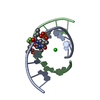
| ||||||||
|---|---|---|---|---|---|---|---|---|---|
| 1 |
| ||||||||
| Unit cell |
|
-Components
| #1: DNA chain | Mass: 3091.049 Da / Num. of mol.: 2 / Source method: obtained synthetically #2: Chemical | ChemComp-SR / |   Mass: 87.620 Da / Num. of mol.: 1 / Source method: obtained synthetically / Formula: Sr Mass: 87.620 Da / Num. of mol.: 1 / Source method: obtained synthetically / Formula: Sr#3: Water | ChemComp-HOH / |  Mass: 18.015 Da / Num. of mol.: 77 / Source method: isolated from a natural source / Formula: H2O Mass: 18.015 Da / Num. of mol.: 77 / Source method: isolated from a natural source / Formula: H2O |
|---|
-Experimental details
-Experiment
| Experiment | Method: X-RAY DIFFRACTION / Number of used crystals: 1 |
|---|
- Sample preparation
| Crystal | Density Matthews: 2.12 Å3/Da / Density % sol: 41.98 % |
|---|---|
| Crystal grow | Temperature: 291 K / Method: vapor diffusion, hanging drop / pH: 7 Details: 40 mM sodium cacodylate, 40 mM lithium chloride, 80 mM strontium chloride, 20 mM magnesium chloride, 12 mM spermine tetrahydrochloride, 10% v/v MPD, pH 7.0, VAPOR DIFFUSION, HANGING DROP, temperature 291K |
-Data collection
| Diffraction | Mean temperature: 100 K |
|---|---|
| Diffraction source | Source: SYNCHROTRON / Site: APS  / Beamline: 21-ID-F / Wavelength: 0.9787 Å / Beamline: 21-ID-F / Wavelength: 0.9787 Å |
| Detector | Type: MARMOSAIC 225 mm CCD / Detector: CCD / Date: Jul 1, 2010 |
| Radiation | Monochromator: C(111) / Protocol: SINGLE WAVELENGTH / Monochromatic (M) / Laue (L): M / Scattering type: x-ray |
| Radiation wavelength | Wavelength: 0.9787 Å / Relative weight: 1 |
| Reflection | Resolution: 1.53→32.45 Å / Num. all: 8268 / Num. obs: 8157 / % possible obs: 98.65 % / Observed criterion σ(F): 0 / Observed criterion σ(I): 0 / Redundancy: 8.4 % / Rmerge(I) obs: 0.07 / Net I/σ(I): 46.3 |
| Reflection shell | Resolution: 1.53→1.57 Å / Redundancy: 8.5 % / Rmerge(I) obs: 0.446 / Mean I/σ(I) obs: 5.7 / % possible all: 70.2 |
- Processing
| Software |
| ||||||||||||||||||||||||||||||||||||||||
|---|---|---|---|---|---|---|---|---|---|---|---|---|---|---|---|---|---|---|---|---|---|---|---|---|---|---|---|---|---|---|---|---|---|---|---|---|---|---|---|---|---|
| Refinement | Method to determine structure: MOLECULAR REPLACEMENT Starting model: PDB ENTRY 3EY2 Resolution: 1.53→32.45 Å / Cor.coef. Fo:Fc: 0.965 / Cor.coef. Fo:Fc free: 0.955 / SU B: 2.963 / SU ML: 0.05 / Cross valid method: THROUGHOUT / σ(F): 0 / σ(I): 0 / ESU R: 0.1 / ESU R Free: 0.09 / Stereochemistry target values: MAXIMUM LIKELIHOOD
| ||||||||||||||||||||||||||||||||||||||||
| Solvent computation | Ion probe radii: 0.8 Å / Shrinkage radii: 0.8 Å / VDW probe radii: 1.4 Å / Solvent model: MASK | ||||||||||||||||||||||||||||||||||||||||
| Displacement parameters | Biso mean: 18.482 Å2
| ||||||||||||||||||||||||||||||||||||||||
| Refinement step | Cycle: LAST / Resolution: 1.53→32.45 Å
| ||||||||||||||||||||||||||||||||||||||||
| Refine LS restraints |
| ||||||||||||||||||||||||||||||||||||||||
| LS refinement shell | Resolution: 1.53→1.569 Å / Total num. of bins used: 20
|
