 Movie
Movie Controller


+ Open data

- Basic information
| Entry | Database: PDB / ID: 3utx | ||||||
|---|---|---|---|---|---|---|---|
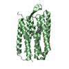
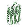
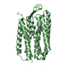
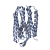
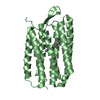
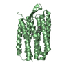
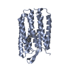
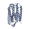
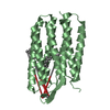
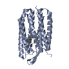
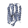
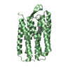
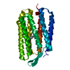
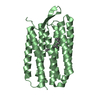
| Title | Crystal structure of bacteriorhodopsin mutant T46A | ||||||
 Components Components | Bacteriorhodopsin | ||||||
 Keywords Keywords | PROTON TRANSPORT / MEMBRANE PROTEIN / PHOTORECEPTOR PROTEIN / RETINAL PROTEIN / ION TRANSPORT / SENSORY TRANSDUCTION | ||||||
| Function / homology |  Function and homology information Function and homology informationlight-driven active monoatomic ion transmembrane transporter activity / phototransduction / photoreceptor activity / monoatomic ion channel activity / proton transmembrane transport / plasma membrane Similarity search - Function | ||||||
| Biological species |  Halobacterium sp. (Halophile) Halobacterium sp. (Halophile) | ||||||
| Method |  X-RAY DIFFRACTION / SYNCHROTRON / MOLECULAR REPLACEMENT / Resolution: 2.47 Å X-RAY DIFFRACTION / SYNCHROTRON / MOLECULAR REPLACEMENT / Resolution: 2.47 Å | ||||||
 Authors Authors | Cao, Z. / Bowie, J.U. | ||||||
 Citation Citation | Journal: Proc.Natl.Acad.Sci.USA / Year: 2012 Title: Shifting hydrogen bonds may produce flexible transmembrane helices. Authors: Cao, Z. / Bowie, J.U. | ||||||
| History |
|
- Structure visualization
| Structure viewer | Molecule: MolmilJmol/JSmol |
|---|
- Downloads & links
-Download
| PDBx/mmCIF format | 3utx.cif.gz | 98.2 KB | Display | PDBx/mmCIF format |
|---|---|---|---|---|
| PDB format | pdb3utx.ent.gz | 75.6 KB | Display | PDB format |
| PDBx/mmJSON format | 3utx.json.gz | Tree view | PDBx/mmJSON format | |
| Others |  Other downloads Other downloads |
-Validation report
| Arichive directory | https://data.pdbj.org/pub/pdb/validation_reports/ut/3utxftp://data.pdbj.org/pub/pdb/validation_reports/ut/3utx | HTTPS FTP |
|---|
-Related structure data


-Links
 PDBj
PDBj


- Assembly
| Deposited unit | 
| ||||||||
|---|---|---|---|---|---|---|---|---|---|
| 1 | 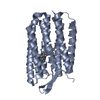
| ||||||||
| 2 | 
| ||||||||
| 3 |
| ||||||||
| Unit cell |
|
-Components
| #1: Protein | Mass: 26899.473 Da / Num. of mol.: 2 / Mutation: T46A Source method: isolated from a genetically manipulated source Source: (gene. exp.) Halobacterium sp. (Halophile) / Strain: ATCC 700922 / JCM 11081 / NRC-1 / Gene: bop, VNG_1467G / Production host: Halobacterium salinarum (Halophile) / Strain (production host): L33 / References: UniProt: P02945#2: Chemical |   Mass: 284.436 Da / Num. of mol.: 2 / Source method: obtained synthetically / Formula: C20H28O Mass: 284.436 Da / Num. of mol.: 2 / Source method: obtained synthetically / Formula: C20H28O#3: Chemical | ChemComp-D12 / |   Mass: 170.335 Da / Num. of mol.: 1 / Source method: obtained synthetically / Formula: C12H26 Mass: 170.335 Da / Num. of mol.: 1 / Source method: obtained synthetically / Formula: C12H26#4: Water | ChemComp-HOH / |  Mass: 18.015 Da / Num. of mol.: 11 / Source method: isolated from a natural source / Formula: H2O Mass: 18.015 Da / Num. of mol.: 11 / Source method: isolated from a natural source / Formula: H2OHas protein modification | Y | |
|---|
-Experimental details
-Experiment
| Experiment | Method: X-RAY DIFFRACTION / Number of used crystals: 1 |
|---|
- Sample preparation
| Crystal | Density Matthews: 2.36 Å3/Da / Density % sol: 47.85 % |
|---|---|
| Crystal grow | Temperature: 310 K / Method: bicelles, vapor diffusion, hanging drop / pH: 4 Details: 0.65M sodium phosphate, 0.95% triethylene glycerol, 0.008M 1,6-hexanediol, 4.3% DMPC, 1.5% CHAPSO, BICELLES, VAPOR DIFFUSION, HANGING DROP, temperature 310K |
-Data collection
| Diffraction | Mean temperature: 100 K |
|---|---|
| Diffraction source | Source: SYNCHROTRON / Site: APS  / Beamline: 24-ID-C / Wavelength: 0.9792 Å / Beamline: 24-ID-C / Wavelength: 0.9792 Å |
| Detector | Type: ADSC QUANTUM 315 / Detector: CCD / Date: Feb 22, 2010 |
| Radiation | Monochromator: Si(111) / Protocol: SINGLE WAVELENGTH / Monochromatic (M) / Laue (L): M / Scattering type: x-ray |
| Radiation wavelength | Wavelength: 0.9792 Å / Relative weight: 1 |
| Reflection | Resolution: 2.47→90 Å / Num. all: 18034 / Num. obs: 18034 / % possible obs: 100 % / Observed criterion σ(F): 0 / Observed criterion σ(I): 0 |
| Reflection shell | Resolution: 2.47→2.56 Å / % possible all: 99.8 |
- Processing
| Software |
| ||||||||||||||||||||
|---|---|---|---|---|---|---|---|---|---|---|---|---|---|---|---|---|---|---|---|---|---|
| Refinement | Method to determine structure: MOLECULAR REPLACEMENT / Resolution: 2.47→51.5 Å / σ(F): 0 / Stereochemistry target values: MAXIMUM LIKELIHOOD
| ||||||||||||||||||||
| Refinement step | Cycle: LAST / Resolution: 2.47→51.5 Å
| ||||||||||||||||||||
| Refine LS restraints |
|
