 Movie
Movie Controller


+ Open data

- Basic information
| Entry | Database: PDB / ID: 3uod | ||||||
|---|---|---|---|---|---|---|---|
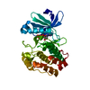
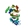
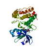
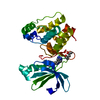
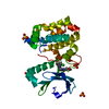
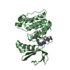
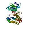
| Title | Aurora A in complex with RPM1693 | ||||||
 Components Components | Aurora kinase A | ||||||
 Keywords Keywords | TRANSFERASE/TRANSFERASE INHIBITOR / protein kinase / aurora A / inhibitor / DFG-in / TRANSFERASE-TRANSFERASE INHIBITOR complex | ||||||
| Function / homology |  Function and homology information Function and homology informationInteraction between PHLDA1 and AURKA / regulation of centrosome cycle / axon hillock / spindle assembly involved in female meiosis I / cilium disassembly / spindle pole centrosome / mitotic centrosome separation / histone H3S10 kinase activity / chromosome passenger complex / positive regulation of oocyte maturation ...Interaction between PHLDA1 and AURKA / regulation of centrosome cycle / axon hillock / spindle assembly involved in female meiosis I / cilium disassembly / spindle pole centrosome / mitotic centrosome separation / histone H3S10 kinase activity / chromosome passenger complex / positive regulation of oocyte maturation / pronucleus / germinal vesicle / protein localization to centrosome / meiotic spindle / anterior/posterior axis specification / neuron projection extension / centrosome localization / spindle organization / positive regulation of mitochondrial fission / mitotic spindle pole / spindle midzone / SUMOylation of DNA replication proteins / negative regulation of protein binding / regulation of G2/M transition of mitotic cell cycle / positive regulation of mitotic cell cycle / positive regulation of mitotic nuclear division / protein serine/threonine/tyrosine kinase activity / centriole / TP53 Regulates Transcription of Genes Involved in G2 Cell Cycle Arrest / liver regeneration / AURKA Activation by TPX2 / molecular function activator activity / regulation of signal transduction by p53 class mediator / mitotic spindle organization / peptidyl-serine phosphorylation / regulation of cytokinesis / regulation of protein stability / response to wounding / APC/C:Cdh1 mediated degradation of Cdc20 and other APC/C:Cdh1 targeted proteins in late mitosis/early G1 / FBXL7 down-regulates AURKA during mitotic entry and in early mitosis / G2/M transition of mitotic cell cycle / kinetochore / spindle / spindle pole / mitotic spindle / Regulation of PLK1 Activity at G2/M Transition / protein autophosphorylation / mitotic cell cycle / positive regulation of proteasomal ubiquitin-dependent protein catabolic process / microtubule cytoskeleton / midbody / Regulation of TP53 Activity through Phosphorylation / basolateral plasma membrane / microtubule / proteasome-mediated ubiquitin-dependent protein catabolic process / protein phosphorylation / protein kinase activity / non-specific serine/threonine protein kinase / ciliary basal body / postsynaptic density / protein heterodimerization activity / negative regulation of gene expression / protein serine kinase activity / cell division / protein serine/threonine kinase activity / apoptotic process / centrosome / ubiquitin protein ligase binding / protein kinase binding / negative regulation of apoptotic process / perinuclear region of cytoplasm / glutamatergic synapse / nucleoplasm / ATP binding / nucleus / cytosol Similarity search - Function | ||||||
| Biological species |  Homo sapiens (human) Homo sapiens (human) | ||||||
| Method |  X-RAY DIFFRACTION / MOLECULAR REPLACEMENT / Resolution: 2.5002 Å X-RAY DIFFRACTION / MOLECULAR REPLACEMENT / Resolution: 2.5002 Å | ||||||
 Authors Authors | Martin, M.P. / Zhu, J.-Y. / Schonbrunn, E. | ||||||
 Citation Citation | Journal: Acs Chem.Biol. / Year: 2012 Title: A Novel Mechanism by Which Small Molecule Inhibitors Induce the DFG Flip in Aurora A. Authors: Martin, M.P. / Zhu, J.Y. / Lawrence, H.R. / Pireddu, R. / Luo, Y. / Alam, R. / Ozcan, S. / Sebti, S.M. / Lawrence, N.J. / Schonbrunn, E. | ||||||
| History |
|
- Structure visualization
| Structure viewer | Molecule: MolmilJmol/JSmol |
|---|
- Downloads & links
-Download
| PDBx/mmCIF format | 3uod.cif.gz | 71.2 KB | Display | PDBx/mmCIF format |
|---|---|---|---|---|
| PDB format | pdb3uod.ent.gz | 51.5 KB | Display | PDB format |
| PDBx/mmJSON format | 3uod.json.gz | Tree view | PDBx/mmJSON format | |
| Others |  Other downloads Other downloads |
-Validation report
| Arichive directory | https://data.pdbj.org/pub/pdb/validation_reports/uo/3uodftp://data.pdbj.org/pub/pdb/validation_reports/uo/3uod | HTTPS FTP |
|---|
-Related structure data
| Related structure data | 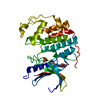 3unjC 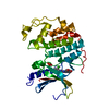 3unkC  3unzC 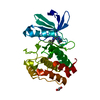 3uo4C 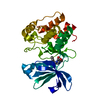 3uo5C 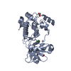 3uo6C  3uohC  3uojC  3uokC 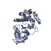 3uolC 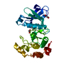 3up2C 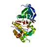 3fdnS C: citing same article ( S: Starting model for refinement |
|---|---|
| Similar structure data |


-Links
 PDBj
PDBj


- Assembly
| Deposited unit | 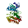
| ||||||||
|---|---|---|---|---|---|---|---|---|---|
| 1 |
| ||||||||
| Unit cell |
|
-Components
| #1: Protein | Mass: 32359.123 Da / Num. of mol.: 1 / Fragment: Kinase domain, RESIDUES 123-401 / Mutation: T287D Source method: isolated from a genetically manipulated source Source: (gene. exp.) Homo sapiens (human)Gene: AURKA, AIK, AIRK1, ARK1, AURA, AYK1, BTAK, IAK1, STK15, STK6 Plasmid: pET28a-MBP / Production host:  References: UniProt: O14965, non-specific serine/threonine protein kinase |
|---|---|
| #2: Chemical | ChemComp-0C3 /   Mass: 374.317 Da / Num. of mol.: 1 / Source method: obtained synthetically / Formula: C18H13F3N4O2 Mass: 374.317 Da / Num. of mol.: 1 / Source method: obtained synthetically / Formula: C18H13F3N4O2 |
| #3: Chemical | ChemComp-EDO /   Mass: 62.068 Da / Num. of mol.: 1 / Source method: obtained synthetically / Formula: C2H6O2 Mass: 62.068 Da / Num. of mol.: 1 / Source method: obtained synthetically / Formula: C2H6O2 |
| #4: Chemical | ChemComp-PEG /   Mass: 106.120 Da / Num. of mol.: 1 / Source method: obtained synthetically / Formula: C4H10O3 Mass: 106.120 Da / Num. of mol.: 1 / Source method: obtained synthetically / Formula: C4H10O3 |
| #5: Water | ChemComp-HOH /  Mass: 18.015 Da / Num. of mol.: 45 / Source method: isolated from a natural source / Formula: H2O Mass: 18.015 Da / Num. of mol.: 45 / Source method: isolated from a natural source / Formula: H2O |
-Experimental details
-Experiment
| Experiment | Method: X-RAY DIFFRACTION / Number of used crystals: 1 |
|---|
- Sample preparation
| Crystal | Density Matthews: 2.61 Å3/Da / Density % sol: 52.79 % |
|---|---|
| Crystal grow | Temperature: 291 K / Method: vapor diffusion, hanging drop / pH: 7.5 Details: 10 mg/mL AURORA A protein, 1 mM RPM1693, 10 % (v/v) PEG 3350, 25 mM phosphate(Na/K pH 7.4), 100 mM sodium tartrate pH 7.0, VAPOR DIFFUSION, HANGING DROP, temperature 291K |
-Data collection
| Diffraction | Mean temperature: 93 K |
|---|---|
| Diffraction source | Source: ROTATING ANODE / Type: RIGAKU MICROMAX-007 HF / Wavelength: 1.54178 / Wavelength: 1.54178 Å |
| Detector | Type: RIGAKU SATURN 944+ / Detector: CCD / Date: Feb 25, 2011 / Details: MIRRORS |
| Radiation | Monochromator: MIRRORS / Protocol: SINGLE WAVELENGTH / Monochromatic (M) / Laue (L): M / Scattering type: x-ray |
| Radiation wavelength | Wavelength: 1.54178 Å / Relative weight: 1 |
| Reflection | Resolution: 2.5→20 Å / Num. obs: 12584 / % possible obs: 99.7 % / Observed criterion σ(I): -3 / Redundancy: 17 % / Rmerge(I) obs: 0.044 / Rsym value: 0.048 / Net I/σ(I): 7.8 |
| Reflection shell | Resolution: 2.5→2.6 Å / Redundancy: 17 % / Rmerge(I) obs: 0.229 / Mean I/σ(I) obs: 7.8 / Rsym value: 0.437 / % possible all: 99.9 |
- Processing
| Software |
| |||||||||||||||||||||||||||||||||||
|---|---|---|---|---|---|---|---|---|---|---|---|---|---|---|---|---|---|---|---|---|---|---|---|---|---|---|---|---|---|---|---|---|---|---|---|---|
| Refinement | Method to determine structure: MOLECULAR REPLACEMENT Starting model: PDB ENTRY 3FDN Resolution: 2.5002→19.691 Å / SU ML: 0.22 / σ(F): 2.01 / Phase error: 23.9 / Stereochemistry target values: ML
| |||||||||||||||||||||||||||||||||||
| Solvent computation | Shrinkage radii: 0.9 Å / VDW probe radii: 1.11 Å / Solvent model: FLAT BULK SOLVENT MODEL / Bsol: 40.655 Å2 / ksol: 0.35 e/Å3 | |||||||||||||||||||||||||||||||||||
| Displacement parameters |
| |||||||||||||||||||||||||||||||||||
| Refine analyze |
| |||||||||||||||||||||||||||||||||||
| Refinement step | Cycle: LAST / Resolution: 2.5002→19.691 Å
| |||||||||||||||||||||||||||||||||||
| Refine LS restraints |
| |||||||||||||||||||||||||||||||||||
| LS refinement shell |
|
