 ムービー
ムービー コントローラー


+ データを開く

- 基本情報
基本情報
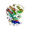
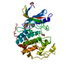
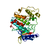
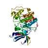
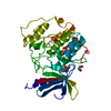
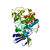
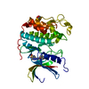
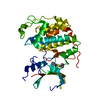
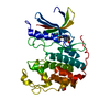
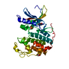
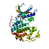
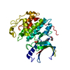
| 登録情報 | データベース: PDB / ID: 3unj | ||||||
|---|---|---|---|---|---|---|---|
| タイトル | CDK2 in complex with inhibitor YL1-038-31 | ||||||
 要素 要素 | Cyclin-dependent kinase 2 | ||||||
 キーワード キーワード | TRANSFERASE/TRANSFERASE INHIBITOR / Protein kinase / allosteric ligand / ANS / TRANSFERASE-TRANSFERASE INHIBITOR complex | ||||||
| 機能・相同性 |  機能・相同性情報 機能・相同性情報cyclin A1-CDK2 complex / cyclin E2-CDK2 complex / cyclin E1-CDK2 complex / cyclin A2-CDK2 complex / positive regulation of DNA-templated DNA replication initiation / cyclin-dependent protein kinase activity / G2 Phase / Y chromosome / Phosphorylation of proteins involved in G1/S transition by active Cyclin E:Cdk2 complexes / positive regulation of heterochromatin formation ...cyclin A1-CDK2 complex / cyclin E2-CDK2 complex / cyclin E1-CDK2 complex / cyclin A2-CDK2 complex / positive regulation of DNA-templated DNA replication initiation / cyclin-dependent protein kinase activity / G2 Phase / Y chromosome / Phosphorylation of proteins involved in G1/S transition by active Cyclin E:Cdk2 complexes / positive regulation of heterochromatin formation / p53-Dependent G1 DNA Damage Response / X chromosome / PTK6 Regulates Cell Cycle / regulation of anaphase-promoting complex-dependent catabolic process / Defective binding of RB1 mutants to E2F1,(E2F2, E2F3) / centriole replication / Regulation of APC/C activators between G1/S and early anaphase / telomere maintenance in response to DNA damage / centrosome duplication / G0 and Early G1 / Telomere Extension By Telomerase / Activation of the pre-replicative complex / cyclin-dependent kinase / cyclin-dependent protein serine/threonine kinase activity / TP53 Regulates Transcription of Genes Involved in G1 Cell Cycle Arrest / Regulation of MITF-M-dependent genes involved in cell cycle and proliferation / Cajal body / Activation of ATR in response to replication stress / Cyclin E associated events during G1/S transition / Cyclin A/B1/B2 associated events during G2/M transition / Cyclin A:Cdk2-associated events at S phase entry / cyclin-dependent protein kinase holoenzyme complex / condensed chromosome / regulation of G2/M transition of mitotic cell cycle / mitotic G1 DNA damage checkpoint signaling / cellular response to nitric oxide / post-translational protein modification / cyclin binding / regulation of mitotic cell cycle / positive regulation of DNA replication / meiotic cell cycle / male germ cell nucleus / CDK-mediated phosphorylation and removal of Cdc6 / G1/S transition of mitotic cell cycle / SCF(Skp2)-mediated degradation of p27/p21 / peptidyl-serine phosphorylation / DNA Damage/Telomere Stress Induced Senescence / Orc1 removal from chromatin / potassium ion transport / Meiotic recombination / Cyclin D associated events in G1 / G2/M transition of mitotic cell cycle / Transcriptional regulation of granulopoiesis / Regulation of TP53 Degradation / cellular senescence / nuclear envelope / Processing of DNA double-strand break ends / regulation of gene expression / Senescence-Associated Secretory Phenotype (SASP) / Factors involved in megakaryocyte development and platelet production / Regulation of TP53 Activity through Phosphorylation / transcription regulator complex / Ras protein signal transduction / chromosome, telomeric region / DNA replication / protein phosphorylation / endosome / chromatin remodeling / protein domain specific binding / cell division / protein serine kinase activity / DNA repair / protein serine/threonine kinase activity / positive regulation of cell population proliferation / DNA-templated transcription / centrosome / positive regulation of DNA-templated transcription / magnesium ion binding / negative regulation of transcription by RNA polymerase II / signal transduction / nucleoplasm / ATP binding / nucleus / cytosol / cytoplasm 類似検索 - 分子機能 | ||||||
| 生物種 |  Homo sapiens (ヒト) Homo sapiens (ヒト) | ||||||
| 手法 |  X線回折 / 分子置換 / 解像度: 1.9001 Å X線回折 / 分子置換 / 解像度: 1.9001 Å | ||||||
 データ登録者 データ登録者 | Zhu, J.-Y. / Martin, M.P. / Alam, R. / Schonbrunn, E. | ||||||
 引用 引用 | ジャーナル: Acs Chem.Biol. / 年: 2012 タイトル: A Novel Mechanism by Which Small Molecule Inhibitors Induce the DFG Flip in Aurora A. 著者: Martin, M.P. / Zhu, J.Y. / Lawrence, H.R. / Pireddu, R. / Luo, Y. / Alam, R. / Ozcan, S. / Sebti, S.M. / Lawrence, N.J. / Schonbrunn, E. | ||||||
| 履歴 |
|
- 構造の表示
構造の表示
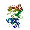
| 構造ビューア | 分子: MolmilJmol/JSmol |
|---|
- ダウンロードとリンク
-ダウンロード
| PDBx/mmCIF形式 | 3unj.cif.gz | 78.2 KB | 表示 | PDBx/mmCIF形式 |
|---|---|---|---|---|
| PDB形式 | pdb3unj.ent.gz | 57.3 KB | 表示 | PDB形式 |
| PDBx/mmJSON形式 | 3unj.json.gz | ツリー表示 | PDBx/mmJSON形式 | |
| その他 |  その他のダウンロード その他のダウンロード |
-検証レポート
| 文書・要旨 | 3unj_validation.pdf.gz | 711 KB | 表示 | wwPDB検証レポート |
|---|---|---|---|---|
| 文書・詳細版 | 3unj_full_validation.pdf.gz | 718.6 KB | 表示 | |
| XML形式データ | 3unj_validation.xml.gz | 15.9 KB | 表示 | |
| CIF形式データ | 3unj_validation.cif.gz | 22.6 KB | 表示 | |
| アーカイブディレクトリ | https://data.pdbj.org/pub/pdb/validation_reports/un/3unjftp://data.pdbj.org/pub/pdb/validation_reports/un/3unj | HTTPS FTP |
-関連構造データ
| 関連構造データ | 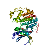 3unkC  3unzC 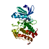 3uo4C 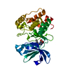 3uo5C  3uo6C 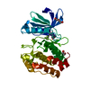 3uodC  3uohC  3uojC  3uokC 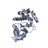 3uolC 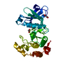 3up2C 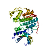 3pxrS C: 同じ文献を引用 ( S: 精密化の開始モデル |
|---|---|
| 類似構造データ |


-リンク
 PDBj
PDBj


- 集合体
集合体
| 登録構造単位 | 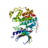
| ||||||||
|---|---|---|---|---|---|---|---|---|---|
| 1 |
| ||||||||
| 単位格子 |
|
-要素
| #1: タンパク質 | 分子量: 33976.488 Da / 分子数: 1 / 由来タイプ: 組換発現 / 由来: (組換発現) Homo sapiens (ヒト) / 遺伝子: CDK2 / プラスミド: pGEX6P-1 / 発現宿主:  |
|---|---|
| #2: 化合物 | ChemComp-0BX / 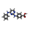  分子量: 306.319 Da / 分子数: 1 / 由来タイプ: 合成 / 式: C17H14N4O2 分子量: 306.319 Da / 分子数: 1 / 由来タイプ: 合成 / 式: C17H14N4O2 |
| #3: 化合物 | ChemComp-PO4 /   分子量: 94.971 Da / 分子数: 1 / 由来タイプ: 合成 / 式: PO4 分子量: 94.971 Da / 分子数: 1 / 由来タイプ: 合成 / 式: PO4 |
| #4: 水 | ChemComp-HOH /  分子量: 18.015 Da / 分子数: 214 / 由来タイプ: 天然 / 式: H2O 分子量: 18.015 Da / 分子数: 214 / 由来タイプ: 天然 / 式: H2O |
-実験情報
-実験
| 実験 | 手法: X線回折 / 使用した結晶の数: 1 |
|---|
- 試料調製
試料調製
| 結晶 | マシュー密度: 2.05 Å3/Da / 溶媒含有率: 39.92 % |
|---|---|
| 結晶化 | 温度: 291 K / 手法: 蒸気拡散法, ハンギングドロップ法 / pH: 7.5 詳細: 5 mg/mL CDK2 protein, 1.5 mM YL1-038-31, 5 % (v/v) PEG 3350, 50 mM HEPES/NaOH (pH 7.5), 50 mM phosphate (Na/K, pH 7.5), VAPOR DIFFUSION, HANGING DROP, temperature 291K |
-データ収集
| 回折 | 平均測定温度: 93 K |
|---|---|
| 放射光源 | 由来: 回転陽極 / タイプ: RIGAKU MICROMAX-007 HF / 波長: 1.54178 / 波長: 1.54178 Å |
| 検出器 | タイプ: RIGAKU RAXIS HTC / 検出器: IMAGE PLATE / 日付: 2009年3月12日 / 詳細: MIRRORS |
| 放射 | モノクロメーター: MIRRORS / プロトコル: SINGLE WAVELENGTH / 単色(M)・ラウエ(L): M / 散乱光タイプ: x-ray |
| 放射波長 | 波長: 1.54178 Å / 相対比: 1 |
| 反射 | 解像度: 1.9→20 Å / Num. obs: 22465 / % possible obs: 99.4 % / Observed criterion σ(I): -3 / 冗長度: 5.7 % / Rmerge(I) obs: 0.059 / Rsym value: 0.059 / Net I/σ(I): 18 |
| 反射 シェル | 解像度: 1.9→2 Å / 冗長度: 5.4 % / Rmerge(I) obs: 0.232 / Mean I/σ(I) obs: 7.5 / Rsym value: 0.254 / % possible all: 99.2 |
- 解析
解析
| ソフトウェア |
| |||||||||||||||||||||||||||||||||||||||||||||||||||||||||||||||
|---|---|---|---|---|---|---|---|---|---|---|---|---|---|---|---|---|---|---|---|---|---|---|---|---|---|---|---|---|---|---|---|---|---|---|---|---|---|---|---|---|---|---|---|---|---|---|---|---|---|---|---|---|---|---|---|---|---|---|---|---|---|---|---|---|
| 精密化 | 構造決定の手法: 分子置換 開始モデル: PDB ENTRY 3PXR 解像度: 1.9001→18.795 Å / SU ML: 0.25 / σ(F): 1.99 / 位相誤差: 25.18 / 立体化学のターゲット値: Engh & Huber
| |||||||||||||||||||||||||||||||||||||||||||||||||||||||||||||||
| 溶媒の処理 | 減衰半径: 0.72 Å / VDWプローブ半径: 1 Å / 溶媒モデル: FLAT BULK SOLVENT MODEL / Bsol: 66.945 Å2 / ksol: 0.383 e/Å3 | |||||||||||||||||||||||||||||||||||||||||||||||||||||||||||||||
| 原子変位パラメータ |
| |||||||||||||||||||||||||||||||||||||||||||||||||||||||||||||||
| Refine analyze |
| |||||||||||||||||||||||||||||||||||||||||||||||||||||||||||||||
| 精密化ステップ | サイクル: LAST / 解像度: 1.9001→18.795 Å
| |||||||||||||||||||||||||||||||||||||||||||||||||||||||||||||||
| 拘束条件 |
| |||||||||||||||||||||||||||||||||||||||||||||||||||||||||||||||
| LS精密化 シェル |
|
