 Movie
Movie Controller


+ Open data

- Basic information
| Entry | Database: PDB / ID: 3ulp | ||||||
|---|---|---|---|---|---|---|---|
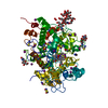
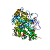
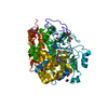
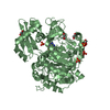
| Title | Plasmodium falciparum SSB complex with ssDNA | ||||||
 Components Components |
| ||||||
 Keywords Keywords | DNA BINDING PROTEIN/DNA / OB-fold / DNA binding / single-stranded DNA / apicoplast / DNA BINDING PROTEIN-DNA complex | ||||||
| Function / homology |  Function and homology information Function and homology informationpositive regulation of mitochondrial DNA replication / apicoplast / : / mitochondrial nucleoid / enzyme activator activity / single-stranded DNA binding / DNA replication Similarity search - Function | ||||||
| Biological species |  Synthetic DNA (others) | ||||||
| Method |  X-RAY DIFFRACTION / MOLECULAR REPLACEMENT / Resolution: 2.1 Å X-RAY DIFFRACTION / MOLECULAR REPLACEMENT / Resolution: 2.1 Å | ||||||
 Authors Authors | Antony, E. / Lohman, T.M. / Korolev, S. | ||||||
 Citation Citation | Journal: J.Mol.Biol. / Year: 2012 Title: Plasmodium falciparum SSB Tetramer Wraps Single-Stranded DNA with Similar Topology but Opposite Polarity to E. coli SSB. Authors: Antony, E. / Weiland, E.A. / Korolev, S. / Lohman, T.M. | ||||||
| History |
|
- Structure visualization
| Structure viewer | Molecule: MolmilJmol/JSmol |
|---|
- Downloads & links
-Download
| PDBx/mmCIF format | 3ulp.cif.gz | 132 KB | Display | PDBx/mmCIF format |
|---|---|---|---|---|
| PDB format | pdb3ulp.ent.gz | 102.9 KB | Display | PDB format |
| PDBx/mmJSON format | 3ulp.json.gz | Tree view | PDBx/mmJSON format | |
| Others |  Other downloads Other downloads |
-Validation report
| Arichive directory | https://data.pdbj.org/pub/pdb/validation_reports/ul/3ulpftp://data.pdbj.org/pub/pdb/validation_reports/ul/3ulp | HTTPS FTP |
|---|
-Related structure data
| Related structure data |  1eqqS S: Starting model for refinement |
|---|---|
| Similar structure data |


-Links
 PDBj
PDBj


- Assembly
| Deposited unit | 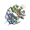
| ||||||||
|---|---|---|---|---|---|---|---|---|---|
| 1 |
| ||||||||
| Unit cell |
| ||||||||
| Components on special symmetry positions |
|
-Components
| #1: Protein | Mass: 14636.719 Da / Num. of mol.: 4 / Fragment: UNP residues 77-200 Source method: isolated from a genetically manipulated source Source: (gene. exp.) Gene: PFE0435c / Production host:  #2: DNA chain | Mass: 10601.791 Da / Num. of mol.: 2 / Source method: obtained synthetically / Source: (synth.) Synthetic DNA (others) #3: Water | ChemComp-HOH / |  Mass: 18.015 Da / Num. of mol.: 228 / Source method: isolated from a natural source / Formula: H2O Mass: 18.015 Da / Num. of mol.: 228 / Source method: isolated from a natural source / Formula: H2O |
|---|
-Experimental details
-Experiment
| Experiment | Method: X-RAY DIFFRACTION / Number of used crystals: 1 |
|---|
- Sample preparation
| Crystal | Density Matthews: 2.64 Å3/Da / Density % sol: 53.49 % |
|---|---|
| Crystal grow | Temperature: 277 K / Method: vapor diffusion, sitting drop / pH: 8.5 Details: 0.1 M Bis-Tris Propane, pH 8.5, 20 % PEG 3350, 0.2 M sodium bromide, VAPOR DIFFUSION, SITTING DROP, temperature 277K |
-Data collection
| Diffraction | Mean temperature: 100 K |
|---|---|
| Diffraction source | Source: ROTATING ANODE / Type: Cu FINE FOCUS / Wavelength: 1.5418 Å |
| Detector | Type: RIGAKU RAXIS IV++ / Detector: IMAGE PLATE / Date: Mar 21, 2011 |
| Radiation | Monochromator: VHF optics / Protocol: SINGLE WAVELENGTH / Monochromatic (M) / Laue (L): M / Scattering type: x-ray |
| Radiation wavelength | Wavelength: 1.5418 Å / Relative weight: 1 |
| Reflection | Resolution: 2.1→50 Å / Num. obs: 48456 / % possible obs: 99.4 % / Observed criterion σ(I): -3 / Redundancy: 3 % / Rmerge(I) obs: 0.068 |
| Reflection shell | Resolution: 2.1→2.14 Å / Redundancy: 3 % / Rmerge(I) obs: 0.529 / Mean I/σ(I) obs: 2.8 / Num. unique all: 2375 / % possible all: 97.3 |
- Processing
| Software |
| |||||||||||||||||||||||||||||||||||||||||||||||||||||||||||||||||
|---|---|---|---|---|---|---|---|---|---|---|---|---|---|---|---|---|---|---|---|---|---|---|---|---|---|---|---|---|---|---|---|---|---|---|---|---|---|---|---|---|---|---|---|---|---|---|---|---|---|---|---|---|---|---|---|---|---|---|---|---|---|---|---|---|---|---|
| Refinement | Method to determine structure: MOLECULAR REPLACEMENT Starting model: PDB ENTRY 1EQQ Resolution: 2.1→30 Å / Cor.coef. Fo:Fc: 0.947 / Cor.coef. Fo:Fc free: 0.911 / SU B: 5.001 / SU ML: 0.133 / Cross valid method: THROUGHOUT / ESU R Free: 0.193 / Stereochemistry target values: MAXIMUM LIKELIHOOD / Details: HYDROGENS HAVE BEEN ADDED IN THE RIDING POSITIONS
| |||||||||||||||||||||||||||||||||||||||||||||||||||||||||||||||||
| Solvent computation | Ion probe radii: 0.8 Å / Shrinkage radii: 0.8 Å / VDW probe radii: 1.4 Å / Solvent model: MASK | |||||||||||||||||||||||||||||||||||||||||||||||||||||||||||||||||
| Displacement parameters | Biso mean: 40.239 Å2
| |||||||||||||||||||||||||||||||||||||||||||||||||||||||||||||||||
| Refinement step | Cycle: LAST / Resolution: 2.1→30 Å
| |||||||||||||||||||||||||||||||||||||||||||||||||||||||||||||||||
| Refine LS restraints |
| |||||||||||||||||||||||||||||||||||||||||||||||||||||||||||||||||
| LS refinement shell | Resolution: 2.1→2.154 Å / Total num. of bins used: 20
|
