 Movie
Movie Controller


+ Open data

- Basic information
| Entry | Database: PDB / ID: 3uc8 | ||||||
|---|---|---|---|---|---|---|---|
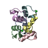
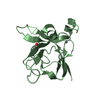
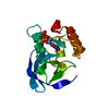
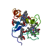
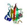
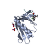
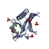
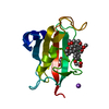
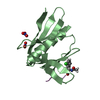
| Title | Trp-cage cyclo-TC1 - tetragonal crystal form | ||||||
 Components Components | cyclo-TC1 | ||||||
 Keywords Keywords | DE NOVO PROTEIN / miniprotein / Trp-cage / cyclic peptide / multimer / protein-protein interaction | ||||||
| Method |  X-RAY DIFFRACTION / MOLECULAR REPLACEMENT / molecular replacement / Resolution: 1.33 Å X-RAY DIFFRACTION / MOLECULAR REPLACEMENT / molecular replacement / Resolution: 1.33 Å | ||||||
 Authors Authors | Scian, M. / Le Trong, I. / Stenkamp, R.E. / Andersen, N.H. | ||||||
 Citation Citation | Journal: Proc.Natl.Acad.Sci.USA / Year: 2012 Title: Crystal and NMR structures of a Trp-cage mini-protein benchmark for computational fold prediction. Authors: Scian, M. / Lin, J.C. / Le Trong, I. / Makhatadze, G.I. / Stenkamp, R.E. / Andersen, N.H. | ||||||
| History |
|
- Structure visualization
| Structure viewer | Molecule:  MolmilJmol/JSmol MolmilJmol/JSmol |
|---|
- Downloads & links
-Download
| PDBx/mmCIF format | 3uc8.cif.gz | 39.6 KB | Display | PDBx/mmCIF format |
|---|---|---|---|---|
| PDB format | pdb3uc8.ent.gz | 29.4 KB | Display | PDB format |
| PDBx/mmJSON format | 3uc8.json.gz | Tree view | PDBx/mmJSON format | |
| Others |  Other downloads Other downloads |
-Validation report
| Arichive directory | https://data.pdbj.org/pub/pdb/validation_reports/uc/3uc8ftp://data.pdbj.org/pub/pdb/validation_reports/uc/3uc8 | HTTPS FTP |
|---|
-Related structure data
| Related structure data | 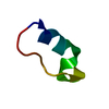 2ll5C  3uc7SC S: Starting model for refinement C: citing same article ( |
|---|---|
| Similar structure data |

-Links
 PDBj
PDBj
- Assembly
| Deposited unit | 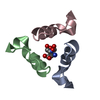
| ||||||||
|---|---|---|---|---|---|---|---|---|---|
| 1 | 
| ||||||||
| Unit cell |
|
-Components
| #1: Protein/peptide | Mass: 2144.240 Da / Num. of mol.: 3 / Source method: obtained synthetically / Details: solid-phase synthesis and cyclization #2: Chemical | ChemComp-TRS / |   Mass: 122.143 Da / Num. of mol.: 1 / Source method: obtained synthetically / Formula: C4H12NO3 / Comment: pH buffer*YM Mass: 122.143 Da / Num. of mol.: 1 / Source method: obtained synthetically / Formula: C4H12NO3 / Comment: pH buffer*YM#3: Water | ChemComp-HOH / |  Mass: 18.015 Da / Num. of mol.: 102 / Source method: isolated from a natural source / Formula: H2O Mass: 18.015 Da / Num. of mol.: 102 / Source method: isolated from a natural source / Formula: H2OHas protein modification | Y | |
|---|
-Experimental details
-Experiment
| Experiment | Method: X-RAY DIFFRACTION / Number of used crystals: 1 |
|---|
- Sample preparation
| Crystal | Density Matthews: 1.75 Å3/Da / Density % sol: 29.59 % |
|---|---|
| Crystal grow | Temperature: 298 K / Method: vapor diffusion, sitting drop / pH: 2.5 Details: 0.2 M Tris-HCl, 0.2 M magnesium chloride, 2.0 M sodium chloride, pH 2.5, VAPOR DIFFUSION, SITTING DROP, temperature 298K |
-Data collection
| Diffraction | Mean temperature: 100 K | |||||||||||||||||||||||||||||||||||||||||||||||||||||||||||||||||||||||||||||
|---|---|---|---|---|---|---|---|---|---|---|---|---|---|---|---|---|---|---|---|---|---|---|---|---|---|---|---|---|---|---|---|---|---|---|---|---|---|---|---|---|---|---|---|---|---|---|---|---|---|---|---|---|---|---|---|---|---|---|---|---|---|---|---|---|---|---|---|---|---|---|---|---|---|---|---|---|---|---|
| Diffraction source | Source: ROTATING ANODE / Type: RIGAKU MICROMAX-007 / Wavelength: 1.5418 Å | |||||||||||||||||||||||||||||||||||||||||||||||||||||||||||||||||||||||||||||
| Detector | Type: RIGAKU RAXIS IV++ / Detector: IMAGE PLATE / Date: Oct 31, 2008 / Details: mirrors | |||||||||||||||||||||||||||||||||||||||||||||||||||||||||||||||||||||||||||||
| Radiation | Protocol: SINGLE WAVELENGTH / Monochromatic (M) / Laue (L): M / Scattering type: x-ray | |||||||||||||||||||||||||||||||||||||||||||||||||||||||||||||||||||||||||||||
| Radiation wavelength | Wavelength: 1.5418 Å / Relative weight: 1 | |||||||||||||||||||||||||||||||||||||||||||||||||||||||||||||||||||||||||||||
| Reflection | Resolution: 1.32→50 Å / Num. all: 9797 / Num. obs: 9797 / % possible obs: 86.2 % / Observed criterion σ(F): 0 / Observed criterion σ(I): 0 / Redundancy: 10 % / Rmerge(I) obs: 0.032 / Χ2: 1.112 / Net I/σ(I): 55.6 | |||||||||||||||||||||||||||||||||||||||||||||||||||||||||||||||||||||||||||||
| Reflection shell |
|
-Phasing
| Phasing | Method: molecular replacement |
|---|
- Processing
| Software |
| ||||||||||||||||||||||||||||||||||||||||||||||||||||||||||||||||||||||||||||||||||||||||||||||||||||
|---|---|---|---|---|---|---|---|---|---|---|---|---|---|---|---|---|---|---|---|---|---|---|---|---|---|---|---|---|---|---|---|---|---|---|---|---|---|---|---|---|---|---|---|---|---|---|---|---|---|---|---|---|---|---|---|---|---|---|---|---|---|---|---|---|---|---|---|---|---|---|---|---|---|---|---|---|---|---|---|---|---|---|---|---|---|---|---|---|---|---|---|---|---|---|---|---|---|---|---|---|---|
| Refinement | Method to determine structure: MOLECULAR REPLACEMENT Starting model: PDB ENTRY 3UC7 Resolution: 1.33→36.85 Å / Cor.coef. Fo:Fc: 0.98 / Cor.coef. Fo:Fc free: 0.956 / WRfactor Rfree: 0.2509 / WRfactor Rwork: 0.1694 / Occupancy max: 1 / Occupancy min: 0.2 / FOM work R set: 0.8821 / SU B: 1.947 / SU ML: 0.035 / SU R Cruickshank DPI: 0.0745 / SU Rfree: 0.0706 / Cross valid method: THROUGHOUT / σ(F): 0 / σ(I): 0 / ESU R: 0.075 / ESU R Free: 0.071 / Stereochemistry target values: MAXIMUM LIKELIHOOD Details: HYDROGENS HAVE BEEN ADDED IN THE RIDING POSITIONS U VALUES : REFINED INDIVIDUALLY
| ||||||||||||||||||||||||||||||||||||||||||||||||||||||||||||||||||||||||||||||||||||||||||||||||||||
| Solvent computation | Ion probe radii: 0.8 Å / Shrinkage radii: 0.8 Å / VDW probe radii: 1.4 Å / Solvent model: MASK | ||||||||||||||||||||||||||||||||||||||||||||||||||||||||||||||||||||||||||||||||||||||||||||||||||||
| Displacement parameters | Biso max: 52.03 Å2 / Biso mean: 15.3019 Å2 / Biso min: 6.14 Å2
| ||||||||||||||||||||||||||||||||||||||||||||||||||||||||||||||||||||||||||||||||||||||||||||||||||||
| Refinement step | Cycle: LAST / Resolution: 1.33→36.85 Å
| ||||||||||||||||||||||||||||||||||||||||||||||||||||||||||||||||||||||||||||||||||||||||||||||||||||
| Refine LS restraints |
| ||||||||||||||||||||||||||||||||||||||||||||||||||||||||||||||||||||||||||||||||||||||||||||||||||||
| LS refinement shell | Resolution: 1.33→1.36 Å / Total num. of bins used: 20
|
