[English] 日本語
Yorodumi
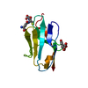
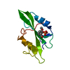
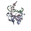
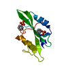
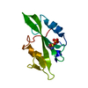
- PDB-3u7z: Crystal structure of a putative metal binding protein RUMGNA_0085... -

+
Open data


ID or keywords:

Loading...

-
Basic information

Entry
Database: PDB / ID: 3u7z
TitleCrystal structure of a putative metal binding protein RUMGNA_00854 (ZP_02040092.1) from Ruminococcus gnavus ATCC 29149 at 1.30 A resolution
Componentsputative metal binding protein RUMGNA_00854
KeywordsMETAL BINDING PROTEIN / THE BINDING PROTEIN / TRANSPORT PROTEIN / STRUCTURAL GENOMICS / JOINT CENTER FOR STRUCTURAL GENOMICS / JCSG / PROTEIN STRUCTURE INITIATIVE / PSI-BIOLOGY
Function / homologyDomain of unknown function DUF4430 / Ferric Hydroxamate Uptake Protein; Chain A, domain 1 - #30 / Domain of unknown function (DUF4430) / Ferric Hydroxamate Uptake Protein; Chain A, domain 1 / Beta Complex / Mainly Beta / metal ion binding / DUF4430 domain-containing protein
Function and homology information
Biological speciesRuminococcus gnavus (bacteria)
MethodX-RAY DIFFRACTION / SYNCHROTRON / MOLECULAR REPLACEMENT / molecular replacement / Resolution: 1.3 Å
AuthorsJoint Center for Structural Genomics (JCSG)
CitationJournal: To be published
Title: Crystal structure of a putative metal binding protein RUMGNA_00854 (ZP_02040092.1) from Ruminococcus gnavus ATCC 29149 at 1.30 A resolution
Authors: Joint Center for Structural Genomics (JCSG)
History
DepositionOct 14, 2011Deposition site: RCSB / Processing site: RCSB
Revision 1.0Nov 2, 2011Provider: repository / Type: Initial release
Revision 1.1Nov 16, 2011Group: Structure summary
Revision 1.2Nov 8, 2017Group: Refinement description / Category: software / Item: _software.classification / _software.name
Revision 1.3Jan 24, 2018Group: Database references / Category: citation_author / Item: _citation_author.name
Revision 1.4Feb 1, 2023Group: Database references / Derived calculations
Category: database_2 / pdbx_struct_conn_angle ...database_2 / pdbx_struct_conn_angle / struct_conn / struct_ref_seq_dif / struct_site
Item: _database_2.pdbx_DOI / _database_2.pdbx_database_accession ..._database_2.pdbx_DOI / _database_2.pdbx_database_accession / _pdbx_struct_conn_angle.ptnr1_auth_asym_id / _pdbx_struct_conn_angle.ptnr1_auth_comp_id / _pdbx_struct_conn_angle.ptnr1_auth_seq_id / _pdbx_struct_conn_angle.ptnr1_label_alt_id / _pdbx_struct_conn_angle.ptnr1_label_asym_id / _pdbx_struct_conn_angle.ptnr1_label_atom_id / _pdbx_struct_conn_angle.ptnr1_label_comp_id / _pdbx_struct_conn_angle.ptnr1_label_seq_id / _pdbx_struct_conn_angle.ptnr2_auth_asym_id / _pdbx_struct_conn_angle.ptnr2_auth_seq_id / _pdbx_struct_conn_angle.ptnr2_label_asym_id / _pdbx_struct_conn_angle.ptnr3_auth_asym_id / _pdbx_struct_conn_angle.ptnr3_auth_comp_id / _pdbx_struct_conn_angle.ptnr3_auth_seq_id / _pdbx_struct_conn_angle.ptnr3_label_alt_id / _pdbx_struct_conn_angle.ptnr3_label_asym_id / _pdbx_struct_conn_angle.ptnr3_label_atom_id / _pdbx_struct_conn_angle.ptnr3_label_comp_id / _pdbx_struct_conn_angle.ptnr3_label_seq_id / _pdbx_struct_conn_angle.value / _struct_conn.pdbx_dist_value / _struct_conn.pdbx_ptnr1_label_alt_id / _struct_conn.pdbx_ptnr2_label_alt_id / _struct_conn.ptnr1_auth_asym_id / _struct_conn.ptnr1_auth_comp_id / _struct_conn.ptnr1_auth_seq_id / _struct_conn.ptnr1_label_asym_id / _struct_conn.ptnr1_label_atom_id / _struct_conn.ptnr1_label_comp_id / _struct_conn.ptnr1_label_seq_id / _struct_conn.ptnr2_auth_asym_id / _struct_conn.ptnr2_auth_comp_id / _struct_conn.ptnr2_auth_seq_id / _struct_conn.ptnr2_label_asym_id / _struct_conn.ptnr2_label_atom_id / _struct_conn.ptnr2_label_comp_id / _struct_conn.ptnr2_label_seq_id / _struct_ref_seq_dif.details / _struct_site.pdbx_auth_asym_id / _struct_site.pdbx_auth_comp_id / _struct_site.pdbx_auth_seq_id
Revision 1.5Sep 20, 2023Group: Data collection / Refinement description
Category: chem_comp_atom / chem_comp_bond / pdbx_initial_refinement_model

-
Structure visualization

Structure viewerMolecule:
MolmilJmol/JSmol

Downloads & links

-
Assembly

Deposited unit
A: putative metal binding protein RUMGNA_00854
B: putative metal binding protein RUMGNA_00854
hetero molecules


Theoretical massNumber of molelcules
Total (without water)23,12722
Polymers22,1492
Non-polymers97720
Water3,009167
1
A: putative metal binding protein RUMGNA_00854
hetero molecules


Theoretical massNumber of molelcules
Total (without water)11,50110
Polymers11,0751
Non-polymers4279
Water181
TypeNameSymmetry operationNumber
identity operation1_555x,y,z1
2
B: putative metal binding protein RUMGNA_00854
hetero molecules


Theoretical massNumber of molelcules
Total (without water)11,62612
Polymers11,0751
Non-polymers55111
Water181
TypeNameSymmetry operationNumber
identity operation1_555x,y,z1
Unit cell
Length a, b, c (Å)29.685, 30.474, 47.156
Angle α, β, γ (deg.)73.130, 83.790, 89.300
Int Tables number1
Space group name H-MP1

-
Components

#1: Protein putative metal binding protein RUMGNA_00854


Mass: 11074.715 Da / Num. of mol.: 2
Source method: isolated from a genetically manipulated source
Source: (gene. exp.) Ruminococcus gnavus (bacteria) / Strain: ATCC 29149 / Gene: RUMGNA_00854 / Plasmid: SpeedET / Production host: Escherichia Coli (E. coli) / Strain (production host): HK100 / References: UniProt: A7AZY1
#2: Chemical
ChemComp-CA / CALCIUM ION


Mass: 40.078 Da / Num. of mol.: 12 / Source method: obtained synthetically / Formula: Ca
#3: Chemical
ChemComp-EDO / 1,2-ETHANEDIOL / ETHYLENE GLYCOL / Ethylene glycol


Mass: 62.068 Da / Num. of mol.: 8 / Source method: obtained synthetically / Formula: C2H6O2
#4: Water ChemComp-HOH / water / Water


Mass: 18.015 Da / Num. of mol.: 167 / Source method: isolated from a natural source / Formula: H2O
Sequence detailsTHE CONSTRUCT (RESIDUES 32-131) WAS EXPRESSED WITH A PURIFICATION TAG MGSDKIHHHHHHENLYFQG. THE TAG ...THE CONSTRUCT (RESIDUES 32-131) WAS EXPRESSED WITH A PURIFICATION TAG MGSDKIHHHHHHENLYFQG. THE TAG WAS REMOVED WITH TEV PROTEASE LEAVING ONLY A GLYCINE (0) FOLLOWED BY THE TARGET SEQUENCE.

-
Experimental details

-
Experiment

ExperimentMethod: X-RAY DIFFRACTION / Number of used crystals: 1

-
Sample preparation

CrystalDensity Matthews: 1.83 Å3/Da / Density % sol: 32.85 %
Crystal growTemperature: 293 K / Method: vapor diffusion, sitting drop / pH: 7.9
Details: 34.0% polyethylene glycol 400, 0.20M calcium acetate, 0.1M HEPES pH 7.9, NANODROP, VAPOR DIFFUSION, SITTING DROP, temperature 293K

-
Data collection

DiffractionMean temperature: 100 K
Diffraction sourceSource: SYNCHROTRON / Site: SSRL / Beamline: BL9-2 / Wavelength: 1
DetectorType: MARMOSAIC 325 mm CCD / Detector: CCD / Date: Jun 23, 2011 / Details: double crystal monochromator
RadiationMonochromator: double crystal / Protocol: SINGLE WAVELENGTH / Monochromatic (M) / Laue (L): M / Scattering type: x-ray
Radiation wavelengthWavelength: 1 Å / Relative weight: 1
ReflectionResolution: 1.3→29.504 Å / Num. all: 36776 / Num. obs: 36776 / % possible obs: 95 % / Redundancy: 2.5 % / Rsym value: 0.067 / Net I/σ(I): 7.9
Reflection shell

Diffraction-ID: 1 / Redundancy: 2.5 %

Resolution (Å)Rmerge(I) obsMean I/σ(I) obsNum. measured allNum. unique allRsym value% possible all
1.3-1.330.5891.2675426540.58992.2
1.33-1.370.4961.4655525810.49692.4
1.37-1.410.4211.7646125400.42193.1
1.41-1.450.3392.1619324400.33993.4
1.45-1.50.2742.6611224100.27493.9
1.5-1.550.2083.5596623520.20894.5
1.55-1.610.1684.2565622200.16894.7
1.61-1.680.1425553421850.14295
1.68-1.750.116.3537421160.1195.7
1.75-1.840.0868508520120.08695.7
1.84-1.940.0729484819090.07296.2
1.94-2.060.0610.4462018240.0696.5
2.06-2.20.05610.9435817210.05697
2.2-2.370.05710.3408716130.05797.3
2.37-2.60.05511.3373914840.05597.6
2.6-2.910.05410.9340013480.05498.1
2.91-3.360.0511.3305112060.0598.4
3.36-4.110.0451224819940.04598
4.11-5.810.0451219147680.04597.1
5.81-29.5040.04712.29833990.04791.2

-
Phasing

PhasingMethod: molecular replacement

-
Processing

Software
NameVersionClassificationNB
MolProbity3beta29model building
PDB_EXTRACT3.1data extraction
MOLREPphasing
SCALA3.3.15data scaling
REFMAC5.5.0110refinement
MOSFLMdata reduction
RefinementMethod to determine structure: MOLECULAR REPLACEMENT
Starting model: PDB entry 2BB6
Resolution: 1.3→29.504 Å / Cor.coef. Fo:Fc: 0.976 / Cor.coef. Fo:Fc free: 0.959 / Occupancy max: 1 / Occupancy min: 0.3 / SU B: 1.805 / SU ML: 0.034 / Cross valid method: THROUGHOUT / σ(F): 0 / ESU R Free: 0.058 / Stereochemistry target values: MAXIMUM LIKELIHOOD
Details: 1. ETHYLENE GLYCOL, CALCIUM MODELED IS PRESENT IN CRYO/CRYSTALLIZATION CONDITIONS. 2. THERE IS SOME UNEXPLAINED,UNUSUAL DENSITY BETWEEN THE CYS100/SG AND LYS128/NZ. 3. HYDROGENS HAVE BEEN ...Details: 1. ETHYLENE GLYCOL, CALCIUM MODELED IS PRESENT IN CRYO/CRYSTALLIZATION CONDITIONS. 2. THERE IS SOME UNEXPLAINED,UNUSUAL DENSITY BETWEEN THE CYS100/SG AND LYS128/NZ. 3. HYDROGENS HAVE BEEN ADDED IN THE RIDING POSITIONS.
RfactorNum. reflection% reflectionSelection details
Rfree0.1887 1824 5 %RANDOM
Rwork0.1379 ---
obs0.1404 36769 95.03 %-
Solvent computationIon probe radii: 0.8 Å / Shrinkage radii: 0.8 Å / VDW probe radii: 1.2 Å / Solvent model: MASK
Displacement parametersBiso max: 50.64 Å2 / Biso mean: 16.8017 Å2 / Biso min: 6.53 Å2
Baniso -1Baniso -2Baniso -3
1-0.02 Å20.13 Å2-0.09 Å2
2--0.06 Å2-0.14 Å2
3---0.01 Å2
Refinement stepCycle: LAST / Resolution: 1.3→29.504 Å
ProteinNucleic acidLigandSolventTotal
Num. atoms1504 0 44 167 1715
Refine LS restraints
Refine-IDTypeDev idealDev ideal targetNumber
X-RAY DIFFRACTIONr_bond_refined_d0.0170.0221655
X-RAY DIFFRACTIONr_bond_other_d0.0020.021060
X-RAY DIFFRACTIONr_angle_refined_deg1.7141.952258
X-RAY DIFFRACTIONr_angle_other_deg1.05332634
X-RAY DIFFRACTIONr_dihedral_angle_1_deg5.9365220
X-RAY DIFFRACTIONr_dihedral_angle_2_deg39.66427.63493
X-RAY DIFFRACTIONr_dihedral_angle_3_deg11.33915275
X-RAY DIFFRACTIONr_chiral_restr0.1060.2250
X-RAY DIFFRACTIONr_gen_planes_refined0.0080.021912
X-RAY DIFFRACTIONr_gen_planes_other0.0010.02296
X-RAY DIFFRACTIONr_mcbond_it2.7313997
X-RAY DIFFRACTIONr_mcbond_other1.6733417
X-RAY DIFFRACTIONr_mcangle_it3.90251629
X-RAY DIFFRACTIONr_scbond_it4.8075658
X-RAY DIFFRACTIONr_scangle_it6.8778615
X-RAY DIFFRACTIONr_rigid_bond_restr2.17732715
X-RAY DIFFRACTIONr_sphericity_free9.4343186
X-RAY DIFFRACTIONr_sphericity_bonded4.22532681
LS refinement shellResolution: 1.3→1.334 Å / Total num. of bins used: 20
RfactorNum. reflection% reflection
Rfree0.333 124 -
Rwork0.231 2526 -
all-2650 -
obs--92.14 %

+
About Yorodumi

-
News

-
Feb 9, 2022. New format data for meta-information of EMDB entries

New format data for meta-information of EMDB entries

  • Version 3 of the EMDB header file is now the official format.
  • The previous official version 1.9 will be removed from the archive.

Related info.:EMDB header

External links:wwPDB to switch to version 3 of the EMDB data model

-
Aug 12, 2020. Covid-19 info

Covid-19 info

URL: https://pdbj.org/emnavi/covid19.php

New page: Covid-19 featured information page in EM Navigator.

Related info.:Covid-19 info / Mar 5, 2020. Novel coronavirus structure data

+
Mar 5, 2020. Novel coronavirus structure data

Novel coronavirus structure data

Related info.:Yorodumi Speices / Aug 12, 2020. Covid-19 info

External links:COVID-19 featured content - PDBj / Molecule of the Month (242):Coronavirus Proteases

+
Jan 31, 2019. EMDB accession codes are about to change! (news from PDBe EMDB page)

EMDB accession codes are about to change! (news from PDBe EMDB page)

  • The allocation of 4 digits for EMDB accession codes will soon come to an end. Whilst these codes will remain in use, new EMDB accession codes will include an additional digit and will expand incrementally as the available range of codes is exhausted. The current 4-digit format prefixed with “EMD-” (i.e. EMD-XXXX) will advance to a 5-digit format (i.e. EMD-XXXXX), and so on. It is currently estimated that the 4-digit codes will be depleted around Spring 2019, at which point the 5-digit format will come into force.
  • The EM Navigator/Yorodumi systems omit the EMD- prefix.

Related info.:Q: What is EMD? / ID/Accession-code notation in Yorodumi/EM Navigator

External links:EMDB Accession Codes are Changing Soon! / Contact to PDBj

+
Jul 12, 2017. Major update of PDB

Major update of PDB

  • wwPDB released updated PDB data conforming to the new PDBx/mmCIF dictionary.
  • This is a major update changing the version number from 4 to 5, and with Remediation, in which all the entries are updated.
  • In this update, many items about electron microscopy experimental information are reorganized (e.g. em_software).
  • Now, EM Navigator and Yorodumi are based on the updated data.

External links:wwPDB Remediation / Enriched Model Files Conforming to OneDep Data Standards Now Available in the PDB FTP Archive

-
Yorodumi

Thousand views of thousand structures

  • Yorodumi is a browser for structure data from EMDB, PDB, SASBDB, etc.
  • This page is also the successor to EM Navigator detail page, and also detail information page/front-end page for Omokage search.
  • The word "yorodu" (or yorozu) is an old Japanese word meaning "ten thousand". "mi" (miru) is to see.

Related info.:EMDB / PDB / SASBDB / Comparison of 3 databanks / Yorodumi Search / Aug 31, 2016. New EM Navigator & Yorodumi / Yorodumi Papers / Jmol/JSmol / Function and homology information / Changes in new EM Navigator and Yorodumi

Read more