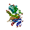
 Movie
Movie Controller

[English] 日本語
 Yorodumi
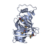
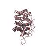
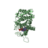
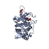
- PDB-3u0i: Crystal Structure of a probable FAD-binding, putative uncharacter... -
+ Open data

- Basic information
| Entry | Database: PDB / ID: 3u0i | ||||||
|---|---|---|---|---|---|---|---|
| Title | Crystal Structure of a probable FAD-binding, putative uncharacterized protein from Brucella melitensis | ||||||
 Components Components | probable FAD-binding, putative uncharacterized protein | ||||||
 Keywords Keywords | UNKNOWN FUNCTION / SSGCID / Emerald BioStructures / Structural Genomics / Seattle Structural Genomics Center for Infectious Disease / FMN-BINDING PROTEIN | ||||||
| Function / homology | Pyridoxamine 5'-phosphate oxidase-related / Pyridoxamine 5'-phosphate oxidase / Electron Transport, Fmn-binding Protein; Chain A / Pnp Oxidase; Chain A / FMN-binding split barrel / Roll / Mainly Beta / Pyridoxamine 5'-phosphate oxidase family protein Function and homology information Function and homology information | ||||||
| Biological species |  Brucella melitensis (bacteria) Brucella melitensis (bacteria) | ||||||
| Method |  X-RAY DIFFRACTION / MOLECULAR REPLACEMENT / molecular replacement / Resolution: 2.2 Å X-RAY DIFFRACTION / MOLECULAR REPLACEMENT / molecular replacement / Resolution: 2.2 Å | ||||||
 Authors Authors | Seattle Structural Genomics Center for Infectious Disease (SSGCID) | ||||||
 Citation Citation | Journal: To be Published Title: Crystal Structure of a probable FAD-binding, putative uncharacterized protein from Brucella melitensis Authors: Seattle Structural Genomics Center for Infectious Disease / Gardberg, A. / Abendroth, J. / Fox, D. / Staker, B. / Stewart, L. | ||||||
| History |
|
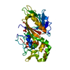
- Structure visualization
| Structure viewer | Molecule: MolmilJmol/JSmol |
|---|
- Downloads & links
-Download
| PDBx/mmCIF format | 3u0i.cif.gz | 68.4 KB | Display | PDBx/mmCIF format |
|---|---|---|---|---|
| PDB format | pdb3u0i.ent.gz | 48.9 KB | Display | PDB format |
| PDBx/mmJSON format | 3u0i.json.gz | Tree view | PDBx/mmJSON format | |
| Others |  Other downloads Other downloads |
-Validation report
| Arichive directory | https://data.pdbj.org/pub/pdb/validation_reports/u0/3u0iftp://data.pdbj.org/pub/pdb/validation_reports/u0/3u0i | HTTPS FTP |
|---|
-Related structure data
| Related structure data |  2hq9S S: Starting model for refinement |
|---|---|
| Similar structure data | |
| Other databases |


-Links
 PDBj
PDBj- Assembly
| Deposited unit | 
| ||||||||
|---|---|---|---|---|---|---|---|---|---|
| 1 | 
| ||||||||
| Unit cell |
|
-Components
| #1: Protein | Mass: 17130.480 Da / Num. of mol.: 1 Source method: isolated from a genetically manipulated source Source: (gene. exp.) Brucella melitensis (bacteria) / Strain: 2308 / Gene: BAB1_1392 / Plasmid: AVA0421 / Production host: |
|---|---|
| #2: Chemical | ChemComp-NHE /   Mass: 207.290 Da / Num. of mol.: 1 / Source method: obtained synthetically / Formula: C8H17NO3S / Comment: pH buffer*YM Mass: 207.290 Da / Num. of mol.: 1 / Source method: obtained synthetically / Formula: C8H17NO3S / Comment: pH buffer*YM |
| #3: Chemical | ChemComp-EDO /   Mass: 62.068 Da / Num. of mol.: 1 / Source method: obtained synthetically / Formula: C2H6O2 Mass: 62.068 Da / Num. of mol.: 1 / Source method: obtained synthetically / Formula: C2H6O2 |
| #4: Water | ChemComp-HOH /  Mass: 18.015 Da / Num. of mol.: 64 / Source method: isolated from a natural source / Formula: H2O Mass: 18.015 Da / Num. of mol.: 64 / Source method: isolated from a natural source / Formula: H2O |
-Experimental details
-Experiment
| Experiment | Method: X-RAY DIFFRACTION / Number of used crystals: 1 |
|---|
- Sample preparation
| Crystal | Density Matthews: 3.32 Å3/Da / Density % sol: 63 % |
|---|---|
| Crystal grow | Temperature: 290 K / Method: vapor diffusion, sitting drop / pH: 9.5 Details: Internal tracking number 222033a7. JCSG screen condition A7: 20% PEG8000, 0.1 M CHES, BrabA.17620.a.A1 PS01056 at 40.2mg/ml, pH 9.5, vapor diffusion, sitting drop, temperature 290K |
-Data collection
| Diffraction | Mean temperature: 100 K | |||||||||||||||||||||||||||||||||||||||||||||||||||||||||||||||||||||||||||||||||||||||||||||||||||||||||||||||||||||||||||||||||||||||||||||||||||
|---|---|---|---|---|---|---|---|---|---|---|---|---|---|---|---|---|---|---|---|---|---|---|---|---|---|---|---|---|---|---|---|---|---|---|---|---|---|---|---|---|---|---|---|---|---|---|---|---|---|---|---|---|---|---|---|---|---|---|---|---|---|---|---|---|---|---|---|---|---|---|---|---|---|---|---|---|---|---|---|---|---|---|---|---|---|---|---|---|---|---|---|---|---|---|---|---|---|---|---|---|---|---|---|---|---|---|---|---|---|---|---|---|---|---|---|---|---|---|---|---|---|---|---|---|---|---|---|---|---|---|---|---|---|---|---|---|---|---|---|---|---|---|---|---|---|---|---|---|
| Diffraction source | Source: ROTATING ANODE / Type: RIGAKU FR-E SUPERBRIGHT / Wavelength: 1.54178 Å | |||||||||||||||||||||||||||||||||||||||||||||||||||||||||||||||||||||||||||||||||||||||||||||||||||||||||||||||||||||||||||||||||||||||||||||||||||
| Detector | Type: RIGAKU SATURN 944+ / Detector: CCD / Date: Sep 27, 2011 | |||||||||||||||||||||||||||||||||||||||||||||||||||||||||||||||||||||||||||||||||||||||||||||||||||||||||||||||||||||||||||||||||||||||||||||||||||
| Radiation | Protocol: SINGLE WAVELENGTH / Monochromatic (M) / Laue (L): M / Scattering type: x-ray | |||||||||||||||||||||||||||||||||||||||||||||||||||||||||||||||||||||||||||||||||||||||||||||||||||||||||||||||||||||||||||||||||||||||||||||||||||
| Radiation wavelength | Wavelength: 1.54178 Å / Relative weight: 1 | |||||||||||||||||||||||||||||||||||||||||||||||||||||||||||||||||||||||||||||||||||||||||||||||||||||||||||||||||||||||||||||||||||||||||||||||||||
| Reflection | Resolution: 2.2→46.21 Å / Num. obs: 21490 / % possible obs: 98.1 % / Observed criterion σ(I): -3 / Redundancy: 3.5 % / Biso Wilson estimate: 43.329 Å2 / Rmerge(I) obs: 0.037 / Net I/σ(I): 23.9 | |||||||||||||||||||||||||||||||||||||||||||||||||||||||||||||||||||||||||||||||||||||||||||||||||||||||||||||||||||||||||||||||||||||||||||||||||||
| Reflection shell | Diffraction-ID: 1
|
-Phasing
| Phasing | Method: molecular replacement |
|---|
- Processing
| Software |
| ||||||||||||||||||||||||||||||||||||||||||||||||||||||||||||
|---|---|---|---|---|---|---|---|---|---|---|---|---|---|---|---|---|---|---|---|---|---|---|---|---|---|---|---|---|---|---|---|---|---|---|---|---|---|---|---|---|---|---|---|---|---|---|---|---|---|---|---|---|---|---|---|---|---|---|---|---|---|
| Refinement | Method to determine structure: MOLECULAR REPLACEMENT Starting model: PDB ENTRY 2HQ9 Resolution: 2.2→46.21 Å / Cor.coef. Fo:Fc: 0.954 / Cor.coef. Fo:Fc free: 0.948 / SU B: 7.486 / SU ML: 0.097 / Cross valid method: THROUGHOUT / σ(F): 0 / ESU R Free: 0.15 / Stereochemistry target values: MAXIMUM LIKELIHOOD Details: U VALUES : WITH TLS ADDED HYDROGENS HAVE BEEN ADDED IN THE RIDING POSITIONS
| ||||||||||||||||||||||||||||||||||||||||||||||||||||||||||||
| Solvent computation | Ion probe radii: 0.8 Å / Shrinkage radii: 0.8 Å / VDW probe radii: 1.2 Å / Solvent model: MASK | ||||||||||||||||||||||||||||||||||||||||||||||||||||||||||||
| Displacement parameters | Biso mean: 37.702 Å2
| ||||||||||||||||||||||||||||||||||||||||||||||||||||||||||||
| Refinement step | Cycle: LAST / Resolution: 2.2→46.21 Å
| ||||||||||||||||||||||||||||||||||||||||||||||||||||||||||||
| Refine LS restraints |
| ||||||||||||||||||||||||||||||||||||||||||||||||||||||||||||
| LS refinement shell | Resolution: 2.2→2.257 Å / Total num. of bins used: 20
| ||||||||||||||||||||||||||||||||||||||||||||||||||||||||||||
| Refinement TLS params. | Method: refined / Origin x: 2.362 Å / Origin y: 56.809 Å / Origin z: 8.039 Å
|
