 Movie
Movie Controller

[English] 日本語
 Yorodumi
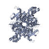
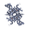
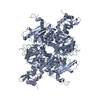
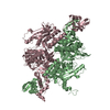
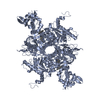
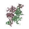
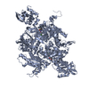
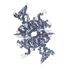
- PDB-3tv5: Crystal Structure of the humanized carboxyltransferase domain of ... -
+ Open data

- Basic information
| Entry | Database: PDB / ID: 3tv5 | ||||||
|---|---|---|---|---|---|---|---|
| Title | Crystal Structure of the humanized carboxyltransferase domain of yeast Acetyl-coA caroxylase in complex with compound 1 | ||||||
 Components Components | Acetyl-CoA carboxylase | ||||||
 Keywords Keywords | LIGASE / carboxyltransferase | ||||||
| Function / homology |  Function and homology information Function and homology informationBiotin transport and metabolism / Fatty acyl-CoA biosynthesis / Carnitine shuttle / acetyl-CoA carboxylase / carboxyl- or carbamoyltransferase activity / acetyl-CoA binding / biotin carboxylase / malonyl-CoA biosynthetic process / acetyl-CoA carboxylase complex / acetyl-CoA biosynthetic process ...Biotin transport and metabolism / Fatty acyl-CoA biosynthesis / Carnitine shuttle / acetyl-CoA carboxylase / carboxyl- or carbamoyltransferase activity / acetyl-CoA binding / biotin carboxylase / malonyl-CoA biosynthetic process / acetyl-CoA carboxylase complex / acetyl-CoA biosynthetic process / biotin carboxylase activity / acetyl-CoA carboxylase activity / long-chain fatty acid biosynthetic process / fatty acid biosynthetic process / protein import into nucleus / endoplasmic reticulum membrane / protein homodimerization activity / mitochondrion / ATP binding / metal ion binding / identical protein binding / cytosol Similarity search - Function | ||||||
| Biological species |  | ||||||
| Method |  X-RAY DIFFRACTION / SYNCHROTRON / MOLECULAR REPLACEMENT / Resolution: 2.8 Å X-RAY DIFFRACTION / SYNCHROTRON / MOLECULAR REPLACEMENT / Resolution: 2.8 Å | ||||||
 Authors Authors | Rajamohan, F. / Marr, E. / Reyes, A. / Landro, J.A. / Anderson, M.D. / Corbett, J.W. / Dirico, K.J. / Harwood, J.H. / Tu, M. / Vajdos, F.F. | ||||||
 Citation Citation | Journal: J.Biol.Chem. / Year: 2011 Title: Structure-guided Inhibitor Design for Human Acetyl-coenzyme A Carboxylase by Interspecies Active Site Conversion. Authors: Rajamohan, F. / Marr, E. / Reyes, A.R. / Landro, J.A. / Anderson, M.D. / Corbett, J.W. / Dirico, K.J. / Harwood, J.H. / Tu, M. / Vajdos, F.F. | ||||||
| History |
|
- Structure visualization
| Structure viewer | Molecule: MolmilJmol/JSmol |
|---|
- Downloads & links
-Download
| PDBx/mmCIF format | 3tv5.cif.gz | 850.7 KB | Display | PDBx/mmCIF format |
|---|---|---|---|---|
| PDB format | pdb3tv5.ent.gz | 707.4 KB | Display | PDB format |
| PDBx/mmJSON format | 3tv5.json.gz | Tree view | PDBx/mmJSON format | |
| Others |  Other downloads Other downloads |
-Validation report
| Arichive directory | https://data.pdbj.org/pub/pdb/validation_reports/tv/3tv5ftp://data.pdbj.org/pub/pdb/validation_reports/tv/3tv5 | HTTPS FTP |
|---|
-Related structure data
| Related structure data |  3tvuC  3tvwC  3tz3C  1w2xS S: Starting model for refinement C: citing same article ( |
|---|---|
| Similar structure data |

-Links
 PDBj
PDBj


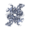
- Assembly
| Deposited unit | 
| ||||||||
|---|---|---|---|---|---|---|---|---|---|
| 1 | 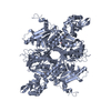
| ||||||||
| 2 | 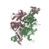
| ||||||||
| Unit cell |
|
-Components
| #1: Protein | Mass: 87220.844 Da / Num. of mol.: 3 Fragment: Carboxyltransferase domain, UNP residues 1476-2233 Mutation: E1919Q,P1920A,H1925F,P1760S,I1762L,M1765V,Q2028E,M2030T,G2032E Source method: isolated from a genetically manipulated source Source: (gene. exp.)  References: UniProt: Q00955, acetyl-CoA carboxylase, biotin carboxylase #2: Chemical | 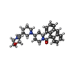  Mass: 485.617 Da / Num. of mol.: 3 / Source method: obtained synthetically / Formula: C30H35N3O3 Mass: 485.617 Da / Num. of mol.: 3 / Source method: obtained synthetically / Formula: C30H35N3O3#3: Water | ChemComp-HOH / |  Mass: 18.015 Da / Num. of mol.: 930 / Source method: isolated from a natural source / Formula: H2O Mass: 18.015 Da / Num. of mol.: 930 / Source method: isolated from a natural source / Formula: H2O |
|---|
-Experimental details
-Experiment
| Experiment | Method: X-RAY DIFFRACTION / Number of used crystals: 1 |
|---|
- Sample preparation
| Crystal | Density Matthews: 4.23 Å3/Da / Density % sol: 70.95 % |
|---|---|
| Crystal grow | Temperature: 298 K / Method: vapor diffusion, hanging drop / pH: 5.5 Details: 100 mM NaCitrate, 12%(w/v) PEG8000, 150 mM LiSO4, 7.5% (v/v) glycerol, pH 5.5, VAPOR DIFFUSION, HANGING DROP, temperature 298K |
-Data collection
| Diffraction | Mean temperature: 100 K |
|---|---|
| Diffraction source | Source: SYNCHROTRON / Site: APS  / Beamline: 17-ID / Wavelength: 1 Å / Beamline: 17-ID / Wavelength: 1 Å |
| Detector | Type: ADSC QUANTUM 210 / Detector: CCD / Date: Jun 11, 2006 |
| Radiation | Protocol: SINGLE WAVELENGTH / Monochromatic (M) / Laue (L): M / Scattering type: x-ray |
| Radiation wavelength | Wavelength: 1 Å / Relative weight: 1 |
| Reflection | Resolution: 2.8→50 Å / Num. obs: 106984 / % possible obs: 99.4 % / Observed criterion σ(I): 2 / Redundancy: 3.4 % / Biso Wilson estimate: 66.78 Å2 / Rmerge(I) obs: 0.089 / Net I/σ(I): 10.3 |
| Reflection shell | Resolution: 2.8→2.9 Å / Redundancy: 3 % / Rmerge(I) obs: 0.386 / % possible all: 97.2 |
- Processing
| Software |
| ||||||||||||||||||||||||||||||||||||||||||||||||||||||||||||||||||||||||||||||||||||||||||||||||||||||||||||||||||
|---|---|---|---|---|---|---|---|---|---|---|---|---|---|---|---|---|---|---|---|---|---|---|---|---|---|---|---|---|---|---|---|---|---|---|---|---|---|---|---|---|---|---|---|---|---|---|---|---|---|---|---|---|---|---|---|---|---|---|---|---|---|---|---|---|---|---|---|---|---|---|---|---|---|---|---|---|---|---|---|---|---|---|---|---|---|---|---|---|---|---|---|---|---|---|---|---|---|---|---|---|---|---|---|---|---|---|---|---|---|---|---|---|---|---|---|
| Refinement | Method to determine structure: MOLECULAR REPLACEMENT Starting model: PDB ENTRY 1W2X Resolution: 2.8→47.06 Å / Cor.coef. Fo:Fc: 0.945 / Cor.coef. Fo:Fc free: 0.9262 / Cross valid method: THROUGHOUT / σ(F): 0
| ||||||||||||||||||||||||||||||||||||||||||||||||||||||||||||||||||||||||||||||||||||||||||||||||||||||||||||||||||
| Displacement parameters | Biso mean: 56.14 Å2
| ||||||||||||||||||||||||||||||||||||||||||||||||||||||||||||||||||||||||||||||||||||||||||||||||||||||||||||||||||
| Refine analyze | Luzzati coordinate error obs: 0.333 Å | ||||||||||||||||||||||||||||||||||||||||||||||||||||||||||||||||||||||||||||||||||||||||||||||||||||||||||||||||||
| Refinement step | Cycle: LAST / Resolution: 2.8→47.06 Å
| ||||||||||||||||||||||||||||||||||||||||||||||||||||||||||||||||||||||||||||||||||||||||||||||||||||||||||||||||||
| Refine LS restraints |
| ||||||||||||||||||||||||||||||||||||||||||||||||||||||||||||||||||||||||||||||||||||||||||||||||||||||||||||||||||
| LS refinement shell | Resolution: 2.8→2.87 Å / Total num. of bins used: 20
| ||||||||||||||||||||||||||||||||||||||||||||||||||||||||||||||||||||||||||||||||||||||||||||||||||||||||||||||||||
| Refinement TLS params. | Method: refined / Refine-ID: X-RAY DIFFRACTION
| ||||||||||||||||||||||||||||||||||||||||||||||||||||||||||||||||||||||||||||||||||||||||||||||||||||||||||||||||||
| Refinement TLS group |
|
