 Movie
Movie Controller

[English] 日本語
 Yorodumi
- PDB-3h0s: Crystal structure of the carboxyltransferase domain of acetyl-coe... -
+ Open data

- Basic information
| Entry | Database: PDB / ID: 3h0s | ||||||
|---|---|---|---|---|---|---|---|
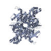
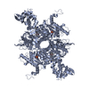
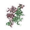
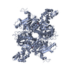
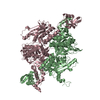
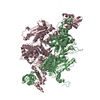
| Title | Crystal structure of the carboxyltransferase domain of acetyl-coenzyme A carboxylase in complex with compound 7 | ||||||
 Components Components | Acetyl-CoA carboxylase | ||||||
 Keywords Keywords | TRANSFERASE / ACETYL-COA CARBOXYLASE / CARBOXYLTRANSFERASE / INHIBITOR / ACC / CT / ATP-binding / Biotin / Cytoplasm / Fatty acid biosynthesis / Ligase / Lipid synthesis / Metal-binding / Multifunctional enzyme / Nucleotide-binding | ||||||
| Function / homology |  Function and homology information Function and homology informationBiotin transport and metabolism / Fatty acyl-CoA biosynthesis / Carnitine shuttle / acetyl-CoA carboxylase / carboxyl- or carbamoyltransferase activity / acetyl-CoA binding / acetyl-CoA carboxylase complex / biotin carboxylase / malonyl-CoA biosynthetic process / acetyl-CoA biosynthetic process ...Biotin transport and metabolism / Fatty acyl-CoA biosynthesis / Carnitine shuttle / acetyl-CoA carboxylase / carboxyl- or carbamoyltransferase activity / acetyl-CoA binding / acetyl-CoA carboxylase complex / biotin carboxylase / malonyl-CoA biosynthetic process / acetyl-CoA biosynthetic process / biotin carboxylase activity / acetyl-CoA carboxylase activity / long-chain fatty acid biosynthetic process / protein import into nucleus / fatty acid biosynthetic process / endoplasmic reticulum membrane / protein homodimerization activity / mitochondrion / ATP binding / metal ion binding / identical protein binding / cytosol Similarity search - Function | ||||||
| Biological species |  | ||||||
| Method |  X-RAY DIFFRACTION / MOLECULAR REPLACEMENT / Resolution: 2.43 Å X-RAY DIFFRACTION / MOLECULAR REPLACEMENT / Resolution: 2.43 Å | ||||||
 Authors Authors | Vajdos, F. | ||||||
 Citation Citation | Journal: Bioorg.Med.Chem.Lett. / Year: 2010 Title: Discovery of small molecule isozyme non-specific inhibitors of mammalian acetyl-CoA carboxylase 1 and 2. Authors: Corbett, J.W. / Freeman-Cook, K.D. / Elliott, R. / Vajdos, F. / Rajamohan, F. / Kohls, D. / Marr, E. / Zhang, H. / Tong, L. / Tu, M. / Murdande, S. / Doran, S.D. / Houser, J.A. / Song, W. / ...Authors: Corbett, J.W. / Freeman-Cook, K.D. / Elliott, R. / Vajdos, F. / Rajamohan, F. / Kohls, D. / Marr, E. / Zhang, H. / Tong, L. / Tu, M. / Murdande, S. / Doran, S.D. / Houser, J.A. / Song, W. / Jones, C.J. / Coffey, S.B. / Buzon, L. / Minich, M.L. / Dirico, K.J. / Tapley, S. / McPherson, R.K. / Sugarman, E. / Harwood, H.J. / Esler, W. | ||||||
| History |
|
- Structure visualization
| Structure viewer | Molecule: MolmilJmol/JSmol |
|---|
- Downloads & links
-Download
| PDBx/mmCIF format | 3h0s.cif.gz | 473.3 KB | Display | PDBx/mmCIF format |
|---|---|---|---|---|
| PDB format | pdb3h0s.ent.gz | 382.8 KB | Display | PDB format |
| PDBx/mmJSON format | 3h0s.json.gz | Tree view | PDBx/mmJSON format | |
| Others |  Other downloads Other downloads |
-Validation report
| Arichive directory | https://data.pdbj.org/pub/pdb/validation_reports/h0/3h0sftp://data.pdbj.org/pub/pdb/validation_reports/h0/3h0s | HTTPS FTP |
|---|
-Related structure data
| Related structure data |  3h0jC  3h0qC  1w2xS C: citing same article ( S: Starting model for refinement |
|---|---|
| Similar structure data |

-Links
 PDBj
PDBj


- Assembly
| Deposited unit | 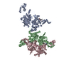
| ||||||||
|---|---|---|---|---|---|---|---|---|---|
| 1 | 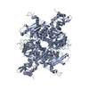
| ||||||||
| 2 | 
| ||||||||
| Unit cell |
|
-Components
| #1: Protein | Mass: 87237.977 Da / Num. of mol.: 3 / Fragment: Residues 1476-2233 Source method: isolated from a genetically manipulated source Source: (gene. exp.) Gene: FAS3, ACC1, YNR016C, N3175 / Production host:  References: UniProt: Q00955, acetyl-CoA carboxylase, biotin carboxylase #2: Chemical |   Mass: 96.063 Da / Num. of mol.: 3 / Source method: obtained synthetically / Formula: SO4 Mass: 96.063 Da / Num. of mol.: 3 / Source method: obtained synthetically / Formula: SO4#3: Chemical |   Mass: 375.420 Da / Num. of mol.: 3 / Source method: obtained synthetically / Formula: C22H21N3O3 Mass: 375.420 Da / Num. of mol.: 3 / Source method: obtained synthetically / Formula: C22H21N3O3#4: Water | ChemComp-HOH / |  Mass: 18.015 Da / Num. of mol.: 2403 / Source method: isolated from a natural source / Formula: H2O Mass: 18.015 Da / Num. of mol.: 2403 / Source method: isolated from a natural source / Formula: H2O |
|---|
-Experimental details
-Experiment
| Experiment | Method: X-RAY DIFFRACTION / Number of used crystals: 1 |
|---|
- Sample preparation
| Crystal | Density Matthews: 4.18 Å3/Da / Density % sol: 70.54 % |
|---|---|
| Crystal grow | Temperature: 298 K / Method: vapor diffusion / pH: 5.5 Details: 0.1 M Na citrate ,pH 5.5, 200 mM NaCl, 8% PEG8000, 10% glycerol, vapor diffusion, temperature 298K |
-Data collection
| Diffraction source | Source: ROTATING ANODE / Type: RIGAKU FR-E SUPERBRIGHT / Wavelength: 1.5418 Å | ||||||||||||||||||||||||||||||||||||||||||||||||||||||||||||||||||
|---|---|---|---|---|---|---|---|---|---|---|---|---|---|---|---|---|---|---|---|---|---|---|---|---|---|---|---|---|---|---|---|---|---|---|---|---|---|---|---|---|---|---|---|---|---|---|---|---|---|---|---|---|---|---|---|---|---|---|---|---|---|---|---|---|---|---|---|
| Detector | Type: RIGAKU RAXIS HTC / Detector: IMAGE PLATE / Date: Jan 16, 2006 | ||||||||||||||||||||||||||||||||||||||||||||||||||||||||||||||||||
| Radiation | Protocol: SINGLE WAVELENGTH / Monochromatic (M) / Laue (L): M / Scattering type: x-ray | ||||||||||||||||||||||||||||||||||||||||||||||||||||||||||||||||||
| Radiation wavelength | Wavelength: 1.5418 Å / Relative weight: 1 | ||||||||||||||||||||||||||||||||||||||||||||||||||||||||||||||||||
| Reflection | Resolution: 2.43→40.7 Å / Num. obs: 159230 / % possible obs: 98.3 % / Redundancy: 3.5 % / Rmerge(I) obs: 0.065 / Χ2: 1.271 / Net I/σ(I): 21.112 | ||||||||||||||||||||||||||||||||||||||||||||||||||||||||||||||||||
| Reflection shell |
|
- Processing
| Software |
| ||||||||||||||||||||||||||||||||||||||||||||||||||||||||||||||||||||||||||||||||||||||||||||||||||||||||||||||||||||||||||||||||||
|---|---|---|---|---|---|---|---|---|---|---|---|---|---|---|---|---|---|---|---|---|---|---|---|---|---|---|---|---|---|---|---|---|---|---|---|---|---|---|---|---|---|---|---|---|---|---|---|---|---|---|---|---|---|---|---|---|---|---|---|---|---|---|---|---|---|---|---|---|---|---|---|---|---|---|---|---|---|---|---|---|---|---|---|---|---|---|---|---|---|---|---|---|---|---|---|---|---|---|---|---|---|---|---|---|---|---|---|---|---|---|---|---|---|---|---|---|---|---|---|---|---|---|---|---|---|---|---|---|---|---|---|
| Refinement | Method to determine structure: MOLECULAR REPLACEMENT Starting model: 1W2X Resolution: 2.43→40.7 Å / Cor.coef. Fo:Fc: 0.967 / Cor.coef. Fo:Fc free: 0.939 / WRfactor Rfree: 0.204 / WRfactor Rwork: 0.148 / Occupancy max: 1 / Occupancy min: 0.35 / FOM work R set: 0.848 / SU B: 6.065 / SU ML: 0.138 / SU R Cruickshank DPI: 0.213 / SU Rfree: 0.2 / Cross valid method: THROUGHOUT / σ(F): 0 / ESU R: 0.213 / ESU R Free: 0.2 / Stereochemistry target values: MAXIMUM LIKELIHOOD / Details: HYDROGENS HAVE BEEN ADDED IN THE RIDING POSITIONS
| ||||||||||||||||||||||||||||||||||||||||||||||||||||||||||||||||||||||||||||||||||||||||||||||||||||||||||||||||||||||||||||||||||
| Solvent computation | Ion probe radii: 0.8 Å / Shrinkage radii: 0.8 Å / VDW probe radii: 1.4 Å / Solvent model: MASK | ||||||||||||||||||||||||||||||||||||||||||||||||||||||||||||||||||||||||||||||||||||||||||||||||||||||||||||||||||||||||||||||||||
| Displacement parameters | Biso max: 164.91 Å2 / Biso mean: 56.371 Å2 / Biso min: 21.96 Å2
| ||||||||||||||||||||||||||||||||||||||||||||||||||||||||||||||||||||||||||||||||||||||||||||||||||||||||||||||||||||||||||||||||||
| Refinement step | Cycle: LAST / Resolution: 2.43→40.7 Å
| ||||||||||||||||||||||||||||||||||||||||||||||||||||||||||||||||||||||||||||||||||||||||||||||||||||||||||||||||||||||||||||||||||
| Refine LS restraints |
| ||||||||||||||||||||||||||||||||||||||||||||||||||||||||||||||||||||||||||||||||||||||||||||||||||||||||||||||||||||||||||||||||||
| LS refinement shell | Resolution: 2.43→2.494 Å / Total num. of bins used: 20
|
