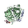
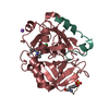
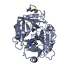
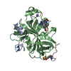
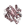
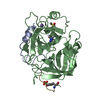
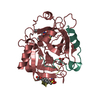
Entry Database : PDB / ID : 3tu7Title Human alpha-thrombin complexed with N-(methylsulfonyl)-D-phenylalanyl-N-((1-carbamimidoyl-4-piperidinyl)methyl)-l-prolinamide (BMS-189664) (Prothrombin) x 2 Hirudin variant-2 Keywords / / Function / homology Function Domain/homology Component
/ / / / / / / / / / / / / / / / / / / / / / / / / / / / / / / / / / / / / / / / / / / / / / / / / / / / / / / / / / / / / / / / / / / / / / / / / / / / / / / / / / / / / / / / / / / / / / / / / / / / / / / / / / / / / / / / / / / / / / / / / / / Biological species Homo sapiens (human)Hirudo medicinalis (medicinal leech)Method / / Resolution : 2.49 Å Authors Malley, M. / Sack, J.S. Journal : Bioorg.Med.Chem.Lett. / Year : 2002Title : Molecular design and structure-activity relationships leading to the potent, selective, and orally active thrombin active site inhibitor BMS-189664.Authors: Das, J. / Kimball, S.D. / Hall, S.E. / Han, W.C. / Iwanowicz, E. / Lin, J. / Moquin, R.V. / Reid, J.A. / Sack, J.S. / Malley, M.F. / Chang, C.Y. / Chong, S. / Wang-Iverson, D.B. / Roberts, D. ... Authors : Das, J. / Kimball, S.D. / Hall, S.E. / Han, W.C. / Iwanowicz, E. / Lin, J. / Moquin, R.V. / Reid, J.A. / Sack, J.S. / Malley, M.F. / Chang, C.Y. / Chong, S. / Wang-Iverson, D.B. / Roberts, D.G. / Seiler, S.M. / Schumacher, W.A. / Ogletree, M.L. History Deposition Sep 16, 2011 Deposition site / Processing site Revision 1.0 Oct 12, 2011 Provider / Type Revision 1.1 Dec 12, 2012 Group Revision 1.2 Nov 20, 2024 Group Data collection / Database references ... Data collection / Database references / Derived calculations / Structure summary Category chem_comp_atom / chem_comp_bond ... chem_comp_atom / chem_comp_bond / database_2 / pdbx_entry_details / pdbx_modification_feature / struct_conn / struct_site Item _database_2.pdbx_DOI / _database_2.pdbx_database_accession ... _database_2.pdbx_DOI / _database_2.pdbx_database_accession / _struct_conn.pdbx_leaving_atom_flag / _struct_site.pdbx_auth_asym_id / _struct_site.pdbx_auth_comp_id / _struct_site.pdbx_auth_seq_id
Show all Show less
 Movie
Movie Controller

 Yorodumi
 Open data
 Basic information
 Components
 Keywords
 Function and homology information
 Homo sapiens (human)
 Hirudo medicinalis (medicinal leech)
 X-RAY DIFFRACTION /
  Authors
 Citation
 Structure visualization
 Downloads & links
 Other downloads
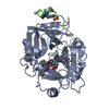
 PDBj
PDBj


 Assembly

 Type: Peptide-like / Class: Thrombin inhibitor / Mass: 478.608 Da / Num. of mol.: 1 / Source method: obtained synthetically / Formula: C22H34N6O4S
 Mass: 18.015 Da / Num. of mol.: 44 / Source method: isolated from a natural source / Formula: H2O
 Sample preparation
 Processing