 Movie
Movie Controller

[English] 日本語
 Yorodumi
- PDB-3ttg: Crystal structure of putative aminomethyltransferase from Leptosp... -
+ Open data

- Basic information
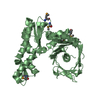
| Entry | Database: PDB / ID: 3ttg | ||||||
|---|---|---|---|---|---|---|---|
| Title | Crystal structure of putative aminomethyltransferase from Leptospirillum rubarum | ||||||
 Components Components | Putative aminomethyltransferase | ||||||
 Keywords Keywords | TRANSFERASE / Structural Genomics / PSI-Biology / Midwest Center for Structural Genomics / MCSG | ||||||
| Function / homology | Probable tRNA modification gtpase trme; domain 1 / Gyrase A; domain 2 / 2-Layer Sandwich / Alpha Beta / :  Function and homology information Function and homology information | ||||||
| Biological species |  Leptospirillum rubarum (bacteria) Leptospirillum rubarum (bacteria) | ||||||
| Method |  X-RAY DIFFRACTION / SYNCHROTRON / SAD / Resolution: 2 Å X-RAY DIFFRACTION / SYNCHROTRON / SAD / Resolution: 2 Å | ||||||
 Authors Authors | Michalska, K. / Xu, X. / Cui, H. / Chin, S. / Savchenko, A. / Joachimiak, A. / Midwest Center for Structural Genomics (MCSG) | ||||||
 Citation Citation | Journal: TO BE PUBLISHED Title: Crystal structure of putative aminomethyltransferase from Leptospirillum rubarum Authors: Michalska, K. / Xu, X. / Cui, H. / Chin, S. / Savchenko, A. / Joachimiak, A. | ||||||
| History |
|
- Structure visualization
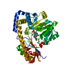
| Structure viewer | Molecule: MolmilJmol/JSmol |
|---|
- Downloads & links
-Download
| PDBx/mmCIF format | 3ttg.cif.gz | 151.8 KB | Display | PDBx/mmCIF format |
|---|---|---|---|---|
| PDB format | pdb3ttg.ent.gz | 119.5 KB | Display | PDB format |
| PDBx/mmJSON format | 3ttg.json.gz | Tree view | PDBx/mmJSON format | |
| Others |  Other downloads Other downloads |
-Validation report
| Arichive directory | https://data.pdbj.org/pub/pdb/validation_reports/tt/3ttgftp://data.pdbj.org/pub/pdb/validation_reports/tt/3ttg | HTTPS FTP |
|---|
-Related structure data
| Similar structure data | |
|---|---|
| Other databases |


-Links
 PDBj
PDBj- Assembly
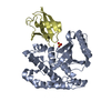
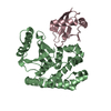
| Deposited unit | 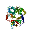
| ||||||||
|---|---|---|---|---|---|---|---|---|---|
| 1 |
| ||||||||
| Unit cell |
| ||||||||
| Components on special symmetry positions |
| ||||||||
| Details | THE AUTHOR STATES THAT THE BIOLOGICAL UNIT OF THIS PROTEIN IS UNKNOWN. |
-Components
| #1: Protein | Mass: 40104.457 Da / Num. of mol.: 1 Source method: isolated from a genetically manipulated source Source: (gene. exp.) Leptospirillum rubarum (bacteria) / Gene: EAY58150, UBAL2_82410567 / Plasmid: p15Tv lic / Production host: | ||||
|---|---|---|---|---|---|
| #2: Chemical | ChemComp-CL /   Mass: 35.453 Da / Num. of mol.: 4 / Source method: obtained synthetically / Formula: Cl Mass: 35.453 Da / Num. of mol.: 4 / Source method: obtained synthetically / Formula: Cl#3: Water | ChemComp-HOH / |  Mass: 18.015 Da / Num. of mol.: 175 / Source method: isolated from a natural source / Formula: H2O Mass: 18.015 Da / Num. of mol.: 175 / Source method: isolated from a natural source / Formula: H2OHas protein modification | Y | |
-Experimental details
-Experiment
| Experiment | Method: X-RAY DIFFRACTION / Number of used crystals: 1 |
|---|
- Sample preparation
| Crystal | Density Matthews: 3.14 Å3/Da / Density % sol: 60.86 % |
|---|---|
| Crystal grow | Temperature: 296 K / pH: 6.9 Details: 0.5 M K3PO4/0.5 M Na3PO4, pH 6.9, VAPOR DIFFUSION, SITTING DROP, temperature 296K |
-Data collection
| Diffraction | Mean temperature: 100 K |
|---|---|
| Diffraction source | Source: SYNCHROTRON / Site: APS  / Beamline: 19-ID / Wavelength: 0.9792 / Beamline: 19-ID / Wavelength: 0.9792 |
| Detector | Type: ADSC QUANTUM 315r / Detector: CCD / Date: Dec 19, 2010 / Details: MIRRORS |
| Radiation | Monochromator: DOUBLE CRYSTAL / Protocol: SINGLE WAVELENGTH / Monochromatic (M) / Laue (L): M / Scattering type: x-ray |
| Radiation wavelength | Wavelength: 0.9792 Å / Relative weight: 1 |
| Reflection | Resolution: 2→50 Å / Num. obs: 34441 / % possible obs: 98.3 % / Observed criterion σ(I): -3 / Redundancy: 11.8 % / Biso Wilson estimate: 36.98 Å2 / Rmerge(I) obs: 0.099 / Net I/σ(I): 23.5 |
| Reflection shell | Resolution: 2→2.03 Å / Redundancy: 5.2 % / Rmerge(I) obs: 0.673 / Mean I/σ(I) obs: 3 / % possible all: 94.4 |
- Processing
| Software |
| |||||||||||||||||||||||||||||||||||||||||||||||||||||||||||||||||||||||||||||||||||||||||||||||||||||||||||||||||||||||||||||||||||||||||||||||||||||||||||||||||||||||||||||||
|---|---|---|---|---|---|---|---|---|---|---|---|---|---|---|---|---|---|---|---|---|---|---|---|---|---|---|---|---|---|---|---|---|---|---|---|---|---|---|---|---|---|---|---|---|---|---|---|---|---|---|---|---|---|---|---|---|---|---|---|---|---|---|---|---|---|---|---|---|---|---|---|---|---|---|---|---|---|---|---|---|---|---|---|---|---|---|---|---|---|---|---|---|---|---|---|---|---|---|---|---|---|---|---|---|---|---|---|---|---|---|---|---|---|---|---|---|---|---|---|---|---|---|---|---|---|---|---|---|---|---|---|---|---|---|---|---|---|---|---|---|---|---|---|---|---|---|---|---|---|---|---|---|---|---|---|---|---|---|---|---|---|---|---|---|---|---|---|---|---|---|---|---|---|---|---|---|
| Refinement | Method to determine structure: SAD / Resolution: 2→36.56 Å / Cor.coef. Fo:Fc: 0.95 / Cor.coef. Fo:Fc free: 0.94 / SU R Cruickshank DPI: 0.179 / Isotropic thermal model: isotropic / Cross valid method: THROUGHOUT / σ(F): 0 / SU R Blow DPI: 0.131 / SU Rfree Blow DPI: 0.124 / SU Rfree Cruickshank DPI: 0.124 / Stereochemistry target values: ML Details: HYDROGEN ATOMS HAVE BEEN ADDED IN THE RIDING POSITIONS
| |||||||||||||||||||||||||||||||||||||||||||||||||||||||||||||||||||||||||||||||||||||||||||||||||||||||||||||||||||||||||||||||||||||||||||||||||||||||||||||||||||||||||||||||
| Displacement parameters | Biso mean: 44.34 Å2
| |||||||||||||||||||||||||||||||||||||||||||||||||||||||||||||||||||||||||||||||||||||||||||||||||||||||||||||||||||||||||||||||||||||||||||||||||||||||||||||||||||||||||||||||
| Refine analyze | Luzzati coordinate error obs: 0.24 Å | |||||||||||||||||||||||||||||||||||||||||||||||||||||||||||||||||||||||||||||||||||||||||||||||||||||||||||||||||||||||||||||||||||||||||||||||||||||||||||||||||||||||||||||||
| Refinement step | Cycle: LAST / Resolution: 2→36.56 Å
| |||||||||||||||||||||||||||||||||||||||||||||||||||||||||||||||||||||||||||||||||||||||||||||||||||||||||||||||||||||||||||||||||||||||||||||||||||||||||||||||||||||||||||||||
| Refine LS restraints |
| |||||||||||||||||||||||||||||||||||||||||||||||||||||||||||||||||||||||||||||||||||||||||||||||||||||||||||||||||||||||||||||||||||||||||||||||||||||||||||||||||||||||||||||||
| LS refinement shell | Resolution: 2→2.06 Å / Total num. of bins used: 17
| |||||||||||||||||||||||||||||||||||||||||||||||||||||||||||||||||||||||||||||||||||||||||||||||||||||||||||||||||||||||||||||||||||||||||||||||||||||||||||||||||||||||||||||||
| Refinement TLS params. | Method: refined / Refine-ID: X-RAY DIFFRACTION
| |||||||||||||||||||||||||||||||||||||||||||||||||||||||||||||||||||||||||||||||||||||||||||||||||||||||||||||||||||||||||||||||||||||||||||||||||||||||||||||||||||||||||||||||
| Refinement TLS group | Selection details: chain A |
