Mass: 18.015 Da / Num. of mol.: 248 / Source method: isolated from a natural source / Formula: H2O
-
Experimental details
-
Experiment
Experiment
Method: X-RAY DIFFRACTION / Number of used crystals: 1
-
Sample preparation
Crystal
Density Matthews: 2.68 Å3/Da / Density % sol: 54.1 %
Crystal grow
Temperature: 290 K / Method: vapor diffusion, sitting drop / pH: 7 Details: Internal tracking number 223543D9. Crystallant (JCSG D9): 25.5% PEG 4000, 15% glycerol, 170 mM Ammonium Sulfate. Protein: BrabA.17351.a.A1 PS01096 at 63 mg/ml in a buffer consisting of 25 mM ...Details: Internal tracking number 223543D9. Crystallant (JCSG D9): 25.5% PEG 4000, 15% glycerol, 170 mM Ammonium Sulfate. Protein: BrabA.17351.a.A1 PS01096 at 63 mg/ml in a buffer consisting of 25 mM HEPES, 300-500 mM NaCl, 2 mM DTT, 0.025% sodium azide, 5% glycerol, pH 7.0, VAPOR DIFFUSION, SITTING DROP, temperature 290K
-
Data collection
Diffraction
Mean temperature: 100 K
Diffraction source
Source: SYNCHROTRON / Site: ALS / Beamline: 5.0.1 / Wavelength: 0.9774 Å
Resolution: 2.15→50 Å / Cor.coef. Fo:Fc: 0.949 / Cor.coef. Fo:Fc free: 0.923 / SU B: 8.276 / SU ML: 0.11 / Cross valid method: THROUGHOUT / σ(F): 0 / ESU R Free: 0.178 / Stereochemistry target values: MAXIMUM LIKELIHOOD Details: U VALUES WITH TLS ADDED. HYDROGENS HAVE BEEN ADDED IN THE RIDING POSITIONS.
Rfactor
Num. reflection
% reflection
Selection details
Rfree
0.228
1751
5 %
RANDOM
Rwork
0.183
-
-
-
obs
0.185
34822
99.89 %
-
Solvent computation
Ion probe radii: 0.8 Å / Shrinkage radii: 0.8 Å / VDW probe radii: 1.2 Å / Solvent model: MASK
Displacement parameters
Biso mean: 26.438 Å2
Baniso -1
Baniso -2
Baniso -3
1-
-0.56 Å2
0 Å2
0 Å2
2-
-
-1.14 Å2
0 Å2
3-
-
-
1.71 Å2
Refinement step
Cycle: LAST / Resolution: 2.15→50 Å
Protein
Nucleic acid
Ligand
Solvent
Total
Num. atoms
3905
0
28
248
4181
Refine LS restraints
Refine-ID
Type
Dev ideal
Dev ideal target
Number
X-RAY DIFFRACTION
r_bond_refined_d
0.012
0.019
4035
X-RAY DIFFRACTION
r_bond_other_d
0.004
0.02
2781
X-RAY DIFFRACTION
r_angle_refined_deg
1.524
1.994
5458
X-RAY DIFFRACTION
r_angle_other_deg
1.166
3
6774
X-RAY DIFFRACTION
r_dihedral_angle_1_deg
5.195
5
533
X-RAY DIFFRACTION
r_dihedral_angle_2_deg
38.019
23.509
171
X-RAY DIFFRACTION
r_dihedral_angle_3_deg
13.068
15
695
X-RAY DIFFRACTION
r_dihedral_angle_4_deg
21.075
15
37
X-RAY DIFFRACTION
r_chiral_restr
0.08
0.2
632
X-RAY DIFFRACTION
r_gen_planes_refined
0.007
0.021
4542
X-RAY DIFFRACTION
r_gen_planes_other
0.004
0.02
815
LS refinement shell
Resolution: 2.15→2.206 Å / Total num. of bins used: 20
Rfactor
Num. reflection
% reflection
Rfree
0.262
97
-
Rwork
0.205
2241
-
all
-
2338
-
obs
-
-
100 %
Refinement TLS params.
Method: refined / Refine-ID: X-RAY DIFFRACTION
ID
L11 (°2)
L12 (°2)
L13 (°2)
L22 (°2)
L23 (°2)
L33 (°2)
S11 (Å °)
S12 (Å °)
S13 (Å °)
S21 (Å °)
S22 (Å °)
S23 (Å °)
S31 (Å °)
S32 (Å °)
S33 (Å °)
T11 (Å2)
T12 (Å2)
T13 (Å2)
T22 (Å2)
T23 (Å2)
T33 (Å2)
Origin x (Å)
Origin y (Å)
Origin z (Å)
1
11.9725
1.233
2.5202
1.2074
0.3457
3.1728
-0.0799
-0.4246
0.439
-0.1126
-0.0836
0.1384
-0.2131
-0.2148
0.1634
0.0381
0.0053
0.0032
0.0337
-0.0505
0.1026
-25.4845
-4.4604
17.2586
2
1.3785
1.0869
0.3704
2.0087
0.3656
0.3339
-0.107
0.1464
-0.0755
-0.1083
0.1246
-0.1448
-0.0113
0.0458
-0.0176
0.0254
-0.023
-0.0007
0.0279
-0.0114
0.0522
5.8139
8.6231
16.0592
3
3.8034
1.2016
-1.329
4.3499
-1.53
3.6238
0.0336
0.2176
0.3239
-0.0305
-0.0122
0.432
-0.0564
-0.0886
-0.0214
0.0367
0.004
-0.0444
0.0307
-0.0096
0.1344
-0.4381
24.9986
22.1562
4
1.9394
-0.1174
-1.3794
2.1898
1.4334
3.6764
-0.1956
0.2557
0.0583
-0.3053
0.1466
-0.1124
-0.2632
-0.0524
0.049
0.0987
-0.0643
-0.0134
0.06
0.0082
0.0842
13.2525
25.5735
15.8393
5
2.2505
3.1589
1.1352
18.7245
-0.3175
3.284
-0.4638
0.2031
0.2737
-0.4638
0.2316
0.3415
-0.2318
-0.0855
0.2322
0.104
-0.0487
-0.0772
0.0368
0.0324
0.1122
-10.349
7.5758
10.3707
6
1.7168
0.7655
0.4811
0.7824
0.1322
0.5671
0.1102
-0.1106
-0.135
0.0563
-0.0446
-0.0944
0.0393
-0.035
-0.0656
0.0256
-0.0178
-0.0123
0.0148
0.0067
0.0237
-26.9068
-21.1564
14.773
7
5.6949
0.0762
-1.8946
2.7406
0.4324
3.1313
-0.039
-0.1473
0.2625
0.157
0.0648
0.0566
-0.0891
-0.043
-0.0258
0.0555
0.002
-0.0289
0.0116
-0.002
0.0334
-41.3421
-15.1478
6.0147
8
1.6702
0.0366
1.0532
0.7715
-0.6665
4.1809
0.1966
-0.2638
-0.1125
0.0701
-0.1339
0.1143
-0.0017
-0.1924
-0.0627
0.0467
-0.0524
-0.006
0.0826
-0.0026
0.0418
-44.4696
-27.1126
13.0366
Refinement TLS group
ID
Refine-ID
Refine TLS-ID
Auth asym-ID
Auth seq-ID
1
X-RAY DIFFRACTION
1
A
4 - 23
2
X-RAY DIFFRACTION
2
A
24 - 172
3
X-RAY DIFFRACTION
3
A
173 - 205
4
X-RAY DIFFRACTION
4
A
206 - 264
5
X-RAY DIFFRACTION
5
B
5 - 23
6
X-RAY DIFFRACTION
6
B
24 - 172
7
X-RAY DIFFRACTION
7
B
173 - 205
8
X-RAY DIFFRACTION
8
B
206 - 264
+
About Yorodumi
-
News
-
Feb 9, 2022. New format data for meta-information of EMDB entries
New format data for meta-information of EMDB entries
Version 3 of the EMDB header file is now the official format.
The previous official version 1.9 will be removed from the archive.
In the structure databanks used in Yorodumi, some data are registered as the other names, "COVID-19 virus" and "2019-nCoV". Here are the details of the virus and the list of structure data.
Jan 31, 2019. EMDB accession codes are about to change! (news from PDBe EMDB page)
EMDB accession codes are about to change! (news from PDBe EMDB page)
The allocation of 4 digits for EMDB accession codes will soon come to an end. Whilst these codes will remain in use, new EMDB accession codes will include an additional digit and will expand incrementally as the available range of codes is exhausted. The current 4-digit format prefixed with “EMD-” (i.e. EMD-XXXX) will advance to a 5-digit format (i.e. EMD-XXXXX), and so on. It is currently estimated that the 4-digit codes will be depleted around Spring 2019, at which point the 5-digit format will come into force.
The EM Navigator/Yorodumi systems omit the EMD- prefix.
Related info.:Q: What is EMD? / ID/Accession-code notation in Yorodumi/EM Navigator
Yorodumi is a browser for structure data from EMDB, PDB, SASBDB, etc.
This page is also the successor to EM Navigator detail page, and also detail information page/front-end page for Omokage search.
The word "yorodu" (or yorozu) is an old Japanese word meaning "ten thousand". "mi" (miru) is to see.
Related info.:EMDB / PDB / SASBDB / Comparison of 3 databanks / Yorodumi Search / Aug 31, 2016. New EM Navigator & Yorodumi / Yorodumi Papers / Jmol/JSmol / Function and homology information / Changes in new EM Navigator and Yorodumi
 Movie
Movie Controller

 Yorodumi
 Open data
 Basic information
 Components
 Keywords
 Function and homology information
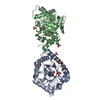
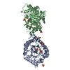
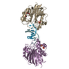
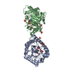
 Brucella melitensis (bacteria)
 X-RAY DIFFRACTION /
  Authors
 Citation
 Structure visualization
 Downloads & links
 Other downloads
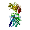

 PDBj
PDBj


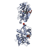
 Assembly

 Mass: 92.094 Da / Num. of mol.: 3 / Source method: obtained synthetically / Formula: C3H8O3
 Mass: 96.063 Da / Num. of mol.: 1 / Source method: obtained synthetically / Formula: SO4
 Mass: 18.015 Da / Num. of mol.: 248 / Source method: isolated from a natural source / Formula: H2O
 Sample preparation
 / Beamline: 5.0.1 / Wavelength: 0.9774 Å
 Processing