 Movie
Movie Controller


+ Open data

- Basic information
| Entry | Database: PDB / ID: 3tsa | ||||||
|---|---|---|---|---|---|---|---|
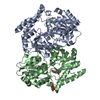
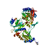
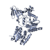
| Title | Spinosyn Rhamnosyltransferase SpnG | ||||||
 Components Components | NDP-rhamnosyltransferase | ||||||
 Keywords Keywords | TRANSFERASE / Glycosyltransferase | ||||||
| Function / homology |  Function and homology information Function and homology informationUDP-glycosyltransferase activity / hexosyltransferase activity / antibiotic biosynthetic process Similarity search - Function | ||||||
| Biological species |  Saccharopolyspora spinosa (bacteria) Saccharopolyspora spinosa (bacteria) | ||||||
| Method |  X-RAY DIFFRACTION / MAD / Resolution: 1.7 Å X-RAY DIFFRACTION / MAD / Resolution: 1.7 Å | ||||||
 Authors Authors | Isiorho, E.A. / Liu, H.-W. / Keatinge-Clay, A.T. | ||||||
 Citation Citation | Journal: Biochemistry / Year: 2012 Title: Structural Studies of the Spinosyn Rhamnosyltransferase, SpnG. Authors: Isiorho, E.A. / Liu, H.W. / Keatinge-Clay, A.T. | ||||||
| History |
|
- Structure visualization
| Structure viewer | Molecule: MolmilJmol/JSmol |
|---|
- Downloads & links
-Download
| PDBx/mmCIF format | 3tsa.cif.gz | 162.5 KB | Display | PDBx/mmCIF format |
|---|---|---|---|---|
| PDB format | pdb3tsa.ent.gz | 126.3 KB | Display | PDB format |
| PDBx/mmJSON format | 3tsa.json.gz | Tree view | PDBx/mmJSON format | |
| Others |  Other downloads Other downloads |
-Validation report
| Arichive directory | https://data.pdbj.org/pub/pdb/validation_reports/ts/3tsaftp://data.pdbj.org/pub/pdb/validation_reports/ts/3tsa | HTTPS FTP |
|---|
-Related structure data
| Related structure data |  3uykC 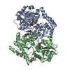 3uylC  2p6pS S: Starting model for refinement C: citing same article ( |
|---|---|
| Similar structure data |


-Links
 PDBj
PDBj

- Assembly
| Deposited unit | 
| ||||||||
|---|---|---|---|---|---|---|---|---|---|
| 1 |
| ||||||||
| Unit cell |
|
-Components
| #1: Protein | Mass: 41356.090 Da / Num. of mol.: 2 Source method: isolated from a genetically manipulated source Source: (gene. exp.) Saccharopolyspora spinosa (bacteria) / Gene: spnG / Production host: References: UniProt: Q9ALM8, Transferases; Glycosyltransferases; Hexosyltransferases #2: Chemical | ChemComp-MG / |   Mass: 24.305 Da / Num. of mol.: 1 / Source method: obtained synthetically / Formula: Mg Mass: 24.305 Da / Num. of mol.: 1 / Source method: obtained synthetically / Formula: Mg#3: Sugar | ChemComp-GLC / |   Type: D-saccharide, alpha linking / Mass: 180.156 Da / Num. of mol.: 1 Type: D-saccharide, alpha linking / Mass: 180.156 Da / Num. of mol.: 1Source method: isolated from a genetically manipulated source Formula: C6H12O6 #4: Water | ChemComp-HOH / |  Mass: 18.015 Da / Num. of mol.: 578 / Source method: isolated from a natural source / Formula: H2O Mass: 18.015 Da / Num. of mol.: 578 / Source method: isolated from a natural source / Formula: H2O |
|---|
-Experimental details
-Experiment
| Experiment | Method: X-RAY DIFFRACTION / Number of used crystals: 1 |
|---|
- Sample preparation
| Crystal | Density Matthews: 2.42 Å3/Da / Density % sol: 49.09 % |
|---|---|
| Crystal grow | Temperature: 298.15 K / Method: vapor diffusion, sitting drop / pH: 6.9 Details: 18% w/v PEG3350, 1% v/v glycerol, 13% w/v glucose, 0.1 M sodium cacodylate, pH 6.9, VAPOR DIFFUSION, SITTING DROP, temperature 298.15K |
-Data collection
| Diffraction | Mean temperature: 100 K |
|---|---|
| Diffraction source | Source: ROTATING ANODE / Type: RIGAKU MICROMAX-007 HF / Wavelength: 1.54 Å |
| Detector | Type: RIGAKU RAXIS IV++ / Detector: IMAGE PLATE / Date: May 31, 2010 |
| Radiation | Monochromator: micromax / Protocol: MAD / Monochromatic (M) / Laue (L): M / Scattering type: x-ray |
| Radiation wavelength | Wavelength: 1.54 Å / Relative weight: 1 |
| Reflection | Resolution: 1.7→19.41 Å / Num. all: 76134 / Num. obs: 72305 / % possible obs: 89.44 % / Observed criterion σ(F): 0 / Observed criterion σ(I): 0 |
| Reflection shell | Resolution: 1.7→1.73 Å / % possible all: 85.8 |
- Processing
| Software |
| ||||||||||||||||||||||||||||||||||||||||||||||||||||||||||||||||||||||||||||||||||||||||||||||||||||||||||||||||||||||||||||||||||||||||||||||||||||||||||||||||||||||||||
|---|---|---|---|---|---|---|---|---|---|---|---|---|---|---|---|---|---|---|---|---|---|---|---|---|---|---|---|---|---|---|---|---|---|---|---|---|---|---|---|---|---|---|---|---|---|---|---|---|---|---|---|---|---|---|---|---|---|---|---|---|---|---|---|---|---|---|---|---|---|---|---|---|---|---|---|---|---|---|---|---|---|---|---|---|---|---|---|---|---|---|---|---|---|---|---|---|---|---|---|---|---|---|---|---|---|---|---|---|---|---|---|---|---|---|---|---|---|---|---|---|---|---|---|---|---|---|---|---|---|---|---|---|---|---|---|---|---|---|---|---|---|---|---|---|---|---|---|---|---|---|---|---|---|---|---|---|---|---|---|---|---|---|---|---|---|---|---|---|---|---|---|
| Refinement | Method to determine structure: MAD Starting model: PDB ENTRY 2P6P Resolution: 1.7→19.37 Å / Cor.coef. Fo:Fc: 0.962 / Cor.coef. Fo:Fc free: 0.937 / SU B: 2.17 / SU ML: 0.072 / Cross valid method: THROUGHOUT / ESU R: 0.117 / ESU R Free: 0.121 / Stereochemistry target values: MAXIMUM LIKELIHOOD / Details: HYDROGENS HAVE BEEN ADDED IN THE RIDING POSITIONS
| ||||||||||||||||||||||||||||||||||||||||||||||||||||||||||||||||||||||||||||||||||||||||||||||||||||||||||||||||||||||||||||||||||||||||||||||||||||||||||||||||||||||||||
| Solvent computation | Ion probe radii: 0.8 Å / Shrinkage radii: 0.8 Å / VDW probe radii: 1.4 Å / Solvent model: MASK | ||||||||||||||||||||||||||||||||||||||||||||||||||||||||||||||||||||||||||||||||||||||||||||||||||||||||||||||||||||||||||||||||||||||||||||||||||||||||||||||||||||||||||
| Displacement parameters | Biso mean: 21.533 Å2
| ||||||||||||||||||||||||||||||||||||||||||||||||||||||||||||||||||||||||||||||||||||||||||||||||||||||||||||||||||||||||||||||||||||||||||||||||||||||||||||||||||||||||||
| Refinement step | Cycle: LAST / Resolution: 1.7→19.37 Å
| ||||||||||||||||||||||||||||||||||||||||||||||||||||||||||||||||||||||||||||||||||||||||||||||||||||||||||||||||||||||||||||||||||||||||||||||||||||||||||||||||||||||||||
| Refine LS restraints |
| ||||||||||||||||||||||||||||||||||||||||||||||||||||||||||||||||||||||||||||||||||||||||||||||||||||||||||||||||||||||||||||||||||||||||||||||||||||||||||||||||||||||||||
| LS refinement shell | Resolution: 1.7→1.744 Å / Total num. of bins used: 20
|
