 Movie
Movie Controller

[English] 日本語
 Yorodumi
- PDB-3tro: Crystal Structure of Leishmania major dihydroorotate dehydrogenas... -
+ Open data

- Basic information
| Entry | Database: PDB / ID: 3tro | ||||||
|---|---|---|---|---|---|---|---|
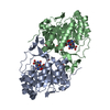
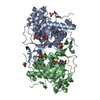
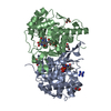
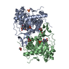
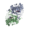
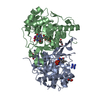
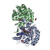
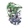
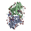
| Title | Crystal Structure of Leishmania major dihydroorotate dehydrogenase mutant D171A | ||||||
 Components Components | Dihydroorotate dehydrogenase | ||||||
 Keywords Keywords | OXIDOREDUCTASE / pyrd / DHODH / LmDHODH | ||||||
| Function / homology |  Function and homology information Function and homology informationdihydroorotate dehydrogenase (fumarate) / dihydroorotate dehydrogenase (fumarate) activity / fumarate metabolic process / dihydroorotate dehydrogenase activity / ciliary plasm / 'de novo' UMP biosynthetic process / 'de novo' pyrimidine nucleobase biosynthetic process / nucleotide binding / nucleoplasm / cytoplasm Similarity search - Function | ||||||
| Biological species |  Leishmania major (eukaryote) Leishmania major (eukaryote) | ||||||
| Method |  X-RAY DIFFRACTION / SYNCHROTRON / MOLECULAR REPLACEMENT / Resolution: 1.86 Å X-RAY DIFFRACTION / SYNCHROTRON / MOLECULAR REPLACEMENT / Resolution: 1.86 Å | ||||||
 Authors Authors | de Souza, A.L. / Pinheiro, M.P. / Nonato, M.C. | ||||||
 Citation Citation | Journal: To be Published Title: Crystal Structure of Leishmania major dihydroorotate dehydrogenase mutant D171A Authors: de Souza, A.L. / Pinheiro, M.P. / Nonato, M.C. | ||||||
| History |
|
- Structure visualization
| Structure viewer | Molecule: MolmilJmol/JSmol |
|---|
- Downloads & links
-Download
| PDBx/mmCIF format | 3tro.cif.gz | 145.9 KB | Display | PDBx/mmCIF format |
|---|---|---|---|---|
| PDB format | pdb3tro.ent.gz | 112.7 KB | Display | PDB format |
| PDBx/mmJSON format | 3tro.json.gz | Tree view | PDBx/mmJSON format | |
| Others |  Other downloads Other downloads |
-Validation report
| Arichive directory | https://data.pdbj.org/pub/pdb/validation_reports/tr/3troftp://data.pdbj.org/pub/pdb/validation_reports/tr/3tro | HTTPS FTP |
|---|
-Related structure data
| Related structure data |  3gyeS  3snq 3snu S: Starting model for refinement |
|---|---|
| Similar structure data |


-Links
 PDBj
PDBj- Assembly
| Deposited unit | 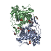
| ||||||||
|---|---|---|---|---|---|---|---|---|---|
| 1 |
| ||||||||
| Unit cell |
|
-Components
| #1: Protein | Mass: 38201.633 Da / Num. of mol.: 2 / Mutation: D171A Source method: isolated from a genetically manipulated source Source: (gene. exp.) Leishmania major (eukaryote) / Strain: Friedlin / Gene: DHODH, lmjf16.0530, LMJF_16_0530 / Plasmid: pET28a / Production host:  #2: Chemical |   Mass: 456.344 Da / Num. of mol.: 2 / Source method: obtained synthetically / Formula: C17H21N4O9P Mass: 456.344 Da / Num. of mol.: 2 / Source method: obtained synthetically / Formula: C17H21N4O9P#3: Chemical |   Mass: 96.063 Da / Num. of mol.: 2 / Source method: obtained synthetically / Formula: SO4 Mass: 96.063 Da / Num. of mol.: 2 / Source method: obtained synthetically / Formula: SO4#4: Chemical | ChemComp-GOL /   Mass: 92.094 Da / Num. of mol.: 8 / Source method: obtained synthetically / Formula: C3H8O3 Mass: 92.094 Da / Num. of mol.: 8 / Source method: obtained synthetically / Formula: C3H8O3#5: Water | ChemComp-HOH / |  Mass: 18.015 Da / Num. of mol.: 433 / Source method: isolated from a natural source / Formula: H2O Mass: 18.015 Da / Num. of mol.: 433 / Source method: isolated from a natural source / Formula: H2O |
|---|
-Experimental details
-Experiment
| Experiment | Method: X-RAY DIFFRACTION / Number of used crystals: 1 |
|---|
- Sample preparation
| Crystal | Density Matthews: 2.71 Å3/Da / Density % sol: 54.63 % |
|---|---|
| Crystal grow | Temperature: 295 K / Method: vapor diffusion, sitting drop / pH: 5.6 Details: 0.1 M sodium citrate tribasic dihydrate, pH 5.6, 1.3 M lithium sulfate, 0.30 M ammonium sulfate , VAPOR DIFFUSION, SITTING DROP, temperature 295K |
-Data collection
| Diffraction | Mean temperature: 100 K |
|---|---|
| Diffraction source | Source: SYNCHROTRON / Site: LNLS  / Beamline: D03B-MX1 / Wavelength: 1.437 Å / Beamline: D03B-MX1 / Wavelength: 1.437 Å |
| Detector | Type: MAR CCD 165 mm / Detector: CCD / Date: Jul 14, 2011 |
| Radiation | Monochromator: Si(111) / Protocol: SINGLE WAVELENGTH / Monochromatic (M) / Laue (L): M / Scattering type: x-ray |
| Radiation wavelength | Wavelength: 1.437 Å / Relative weight: 1 |
| Reflection | Resolution: 1.86→20.82 Å / Num. obs: 68190 / % possible obs: 99.8 % / Observed criterion σ(I): 2.5 / Redundancy: 8 % / Biso Wilson estimate: 16.171 Å2 / Rmerge(I) obs: 0.131 / Rsym value: 0.131 / Net I/σ(I): 14.2 |
| Reflection shell | Resolution: 1.86→1.96 Å / Redundancy: 5.2 % / Rmerge(I) obs: 0.639 / Mean I/σ(I) obs: 2.9 / Num. unique all: 9824 / Rsym value: 0.639 / % possible all: 98.9 |
- Processing
| Software |
| |||||||||||||||||||||||||||||||||||||||||||||
|---|---|---|---|---|---|---|---|---|---|---|---|---|---|---|---|---|---|---|---|---|---|---|---|---|---|---|---|---|---|---|---|---|---|---|---|---|---|---|---|---|---|---|---|---|---|---|
| Refinement | Method to determine structure: MOLECULAR REPLACEMENT Starting model: PDB ENTRY 3GYE Resolution: 1.86→19.61 Å / Cor.coef. Fo:Fc: 0.957 / Cor.coef. Fo:Fc free: 0.936 / SU B: 2.978 / SU ML: 0.085 / Cross valid method: THROUGHOUT / ESU R: 0.119 / ESU R Free: 0.12 / Stereochemistry target values: MAXIMUM LIKELIHOOD Details: HYDROGENS HAVE BEEN USED IF PRESENT IN THE INPUT U VALUES : REFINED INDIVIDUALLY
| |||||||||||||||||||||||||||||||||||||||||||||
| Solvent computation | Ion probe radii: 0.8 Å / Shrinkage radii: 0.8 Å / VDW probe radii: 1.2 Å / Solvent model: MASK | |||||||||||||||||||||||||||||||||||||||||||||
| Displacement parameters | Biso mean: 21.497 Å2
| |||||||||||||||||||||||||||||||||||||||||||||
| Refinement step | Cycle: LAST / Resolution: 1.86→19.61 Å
| |||||||||||||||||||||||||||||||||||||||||||||
| Refine LS restraints |
| |||||||||||||||||||||||||||||||||||||||||||||
| LS refinement shell | Resolution: 1.86→1.912 Å / Total num. of bins used: 20
|
