 Movie
Movie Controller

[English] 日本語
 Yorodumi
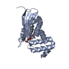
- PDB-3tl1: Crystal structure of the Streptomyces coelicolor WhiE ORFVI polyk... -
+ Open data

- Basic information
| Entry | Database: PDB / ID: 3tl1 | ||||||
|---|---|---|---|---|---|---|---|
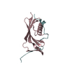
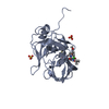
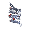
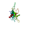
| Title | Crystal structure of the Streptomyces coelicolor WhiE ORFVI polyketide aromatase/cyclase | ||||||
 Components Components | Polyketide cyclase | ||||||
 Keywords Keywords | BIOSYNTHETIC PROTEIN / Helix-grip fold / polyketide C9-C14 aromatase/cyclase / linear poly-beta-ketone intermediate | ||||||
| Function / homology | Coenzyme Q-binding protein COQ10, START domain / Polyketide cyclase / dehydrase and lipid transport / START domain / Alpha-D-Glucose-1,6-Bisphosphate; Chain A, domain 4 / START-like domain superfamily / 2-Layer Sandwich / Alpha Beta / Chem-JRO / Putative polyketide cyclase Function and homology information Function and homology information | ||||||
| Biological species |  Streptomyces coelicolor (bacteria) Streptomyces coelicolor (bacteria) | ||||||
| Method |  X-RAY DIFFRACTION / SYNCHROTRON / MOLECULAR REPLACEMENT / Resolution: 1.8 Å X-RAY DIFFRACTION / SYNCHROTRON / MOLECULAR REPLACEMENT / Resolution: 1.8 Å | ||||||
 Authors Authors | Lee, M.-Y. / Ames, B.D. / Zhang, W. / Tang, Y. / Tsai, S.-C. | ||||||
 Citation Citation | Journal: Biochemistry / Year: 2012 Title: Insight into the Molecular Basis of Aromatic Polyketide Cyclization: Crystal Structure and in Vitro Characterization of WhiE-ORFVI. Authors: Lee, M.Y. / Ames, B.D. / Tsai, S.C. | ||||||
| History |
|
- Structure visualization
| Structure viewer | Molecule: MolmilJmol/JSmol |
|---|
- Downloads & links
-Download
| PDBx/mmCIF format | 3tl1.cif.gz | 144.7 KB | Display | PDBx/mmCIF format |
|---|---|---|---|---|
| PDB format | pdb3tl1.ent.gz | 114.8 KB | Display | PDB format |
| PDBx/mmJSON format | 3tl1.json.gz | Tree view | PDBx/mmJSON format | |
| Others |  Other downloads Other downloads |
-Validation report
| Arichive directory | https://data.pdbj.org/pub/pdb/validation_reports/tl/3tl1ftp://data.pdbj.org/pub/pdb/validation_reports/tl/3tl1 | HTTPS FTP |
|---|
-Related structure data


-Links
 PDBj
PDBj- Assembly
| Deposited unit | 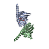
| ||||||||
|---|---|---|---|---|---|---|---|---|---|
| 1 | 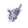
| ||||||||
| 2 | 
| ||||||||
| Unit cell |
|
-Components
| #1: Protein | Mass: 18216.340 Da / Num. of mol.: 2 Source method: isolated from a genetically manipulated source Source: (gene. exp.) Streptomyces coelicolor (bacteria) / Gene: SCO5315, SC6G9.18 / Plasmid: pET28 / Production host: #2: Chemical |   Mass: 258.226 Da / Num. of mol.: 2 / Source method: obtained synthetically / Formula: C14H10O5 Mass: 258.226 Da / Num. of mol.: 2 / Source method: obtained synthetically / Formula: C14H10O5#3: Chemical |   Mass: 92.094 Da / Num. of mol.: 3 / Source method: obtained synthetically / Formula: C3H8O3 Mass: 92.094 Da / Num. of mol.: 3 / Source method: obtained synthetically / Formula: C3H8O3#4: Water | ChemComp-HOH / |  Mass: 18.015 Da / Num. of mol.: 166 / Source method: isolated from a natural source / Formula: H2O Mass: 18.015 Da / Num. of mol.: 166 / Source method: isolated from a natural source / Formula: H2O |
|---|
-Experimental details
-Experiment
| Experiment | Method: X-RAY DIFFRACTION / Number of used crystals: 1 |
|---|
- Sample preparation
| Crystal | Density Matthews: 2.34 Å3/Da / Density % sol: 47.49 % |
|---|---|
| Crystal grow | Temperature: 277 K / Method: vapor diffusion, hanging drop / pH: 5.4 Details: 0.1M Bis-Tris pH5.4, 0.2M magnesium chloride, 30% PEG3350, 5mM JRSi284 (ligand), VAPOR DIFFUSION, HANGING DROP, temperature 277K |
-Data collection
| Diffraction | Mean temperature: 298 K |
|---|---|
| Diffraction source | Source: SYNCHROTRON / Site: ALS  / Beamline: 8.2.2 / Wavelength: 1 Å / Beamline: 8.2.2 / Wavelength: 1 Å |
| Detector | Type: ADSC QUANTUM 315r / Detector: CCD / Date: Feb 24, 2009 |
| Radiation | Monochromator: Double crystal, Si (111) / Protocol: SINGLE WAVELENGTH / Monochromatic (M) / Laue (L): M / Scattering type: x-ray |
| Radiation wavelength | Wavelength: 1 Å / Relative weight: 1 |
| Reflection | Resolution: 1.8→65.37 Å / Num. obs: 31201 / % possible obs: 99.9 % / Observed criterion σ(F): 0 / Observed criterion σ(I): -3 |
| Reflection shell | Resolution: 1.8→1.86 Å / % possible all: 99.9 |
- Processing
| Software |
| ||||||||||||||||||||||||||||||||||||||||||||||||||||||||||||||||||||||||||||||||||||||||||||||||||||||||||||||||||||||||||||||||||||||||||||||||||||||||||||||||||||||||||
|---|---|---|---|---|---|---|---|---|---|---|---|---|---|---|---|---|---|---|---|---|---|---|---|---|---|---|---|---|---|---|---|---|---|---|---|---|---|---|---|---|---|---|---|---|---|---|---|---|---|---|---|---|---|---|---|---|---|---|---|---|---|---|---|---|---|---|---|---|---|---|---|---|---|---|---|---|---|---|---|---|---|---|---|---|---|---|---|---|---|---|---|---|---|---|---|---|---|---|---|---|---|---|---|---|---|---|---|---|---|---|---|---|---|---|---|---|---|---|---|---|---|---|---|---|---|---|---|---|---|---|---|---|---|---|---|---|---|---|---|---|---|---|---|---|---|---|---|---|---|---|---|---|---|---|---|---|---|---|---|---|---|---|---|---|---|---|---|---|---|---|---|
| Refinement | Method to determine structure: MOLECULAR REPLACEMENT / Resolution: 1.8→41.29 Å / Cor.coef. Fo:Fc: 0.967 / Cor.coef. Fo:Fc free: 0.936 / SU B: 7.028 / SU ML: 0.099 / Cross valid method: THROUGHOUT / σ(F): 0 / ESU R Free: 0.149 / Stereochemistry target values: MAXIMUM LIKELIHOOD / Details: HYDROGENS HAVE BEEN ADDED IN THE RIDING POSITIONS
| ||||||||||||||||||||||||||||||||||||||||||||||||||||||||||||||||||||||||||||||||||||||||||||||||||||||||||||||||||||||||||||||||||||||||||||||||||||||||||||||||||||||||||
| Solvent computation | Ion probe radii: 0.8 Å / Shrinkage radii: 0.8 Å / VDW probe radii: 1.2 Å / Solvent model: MASK | ||||||||||||||||||||||||||||||||||||||||||||||||||||||||||||||||||||||||||||||||||||||||||||||||||||||||||||||||||||||||||||||||||||||||||||||||||||||||||||||||||||||||||
| Displacement parameters | Biso mean: 28.67 Å2
| ||||||||||||||||||||||||||||||||||||||||||||||||||||||||||||||||||||||||||||||||||||||||||||||||||||||||||||||||||||||||||||||||||||||||||||||||||||||||||||||||||||||||||
| Refinement step | Cycle: LAST / Resolution: 1.8→41.29 Å
| ||||||||||||||||||||||||||||||||||||||||||||||||||||||||||||||||||||||||||||||||||||||||||||||||||||||||||||||||||||||||||||||||||||||||||||||||||||||||||||||||||||||||||
| Refine LS restraints |
| ||||||||||||||||||||||||||||||||||||||||||||||||||||||||||||||||||||||||||||||||||||||||||||||||||||||||||||||||||||||||||||||||||||||||||||||||||||||||||||||||||||||||||
| LS refinement shell | Resolution: 1.8→1.848 Å / Total num. of bins used: 20
|
