[English] 日本語
Yorodumi
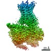
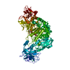
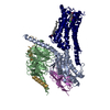
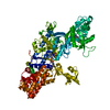
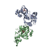
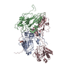
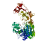
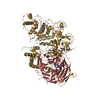
- PDB-3tho: Crystal structure of Mre11:Rad50 in its ATP/ADP bound state -

+
Open data


ID or keywords:

Loading...

-
Basic information

Entry
Database: PDB / ID: 3tho
TitleCrystal structure of Mre11:Rad50 in its ATP/ADP bound state
Components
  • Exonuclease, putative
  • Probable DNA double-strand break repair Rad50 ATPase
KeywordsHYDROLASE/DNA BINDING PROTEIN / Adenosine Triphosphate / Bacterial Proteins / DNA Breaks / Double-Stranded / DNA Repair / DNA Repair Enzymes / DNA-Binding Proteins / Endodeoxyribonucleases / Exodeoxyribonucleases / Models / Molecular / Scattering / Small Angle / Thermotoga maritima / ABC ATPase / nuclease / HYDROLASE / HYDROLASE-DNA BINDING PROTEIN complex
Function / homology
Function and homology information


DNA exonuclease activity / Hydrolases; Acting on acid anhydrides / 3'-5' exonuclease activity / DNA endonuclease activity / double-strand break repair / DNA recombination / DNA replication / Hydrolases; Acting on ester bonds / DNA repair / ATP hydrolysis activity ...DNA exonuclease activity / Hydrolases; Acting on acid anhydrides / 3'-5' exonuclease activity / DNA endonuclease activity / double-strand break repair / DNA recombination / DNA replication / Hydrolases; Acting on ester bonds / DNA repair / ATP hydrolysis activity / DNA binding / ATP binding / metal ion binding
Similarity search - Function
Helix Hairpins - #980 / DNA double-strand break repair nuclease / Helix hairpin bin / RAD50, zinc hook / Rad50 zinc-hook domain profile. / Nuclease SbcCD subunit D / : / SbcC/RAD50-like, Walker B motif / Mre11 nuclease, N-terminal metallophosphatase domain / Rad50/SbcC-type AAA domain ...Helix Hairpins - #980 / DNA double-strand break repair nuclease / Helix hairpin bin / RAD50, zinc hook / Rad50 zinc-hook domain profile. / Nuclease SbcCD subunit D / : / SbcC/RAD50-like, Walker B motif / Mre11 nuclease, N-terminal metallophosphatase domain / Rad50/SbcC-type AAA domain / AAA domain / Helix hairpin bin domain superfamily / Metallo-dependent phosphatases / Purple Acid Phosphatase; chain A, domain 2 / Calcineurin-like phosphoesterase domain, ApaH type / Calcineurin-like phosphoesterase / Helix Hairpins / Metallo-dependent phosphatase-like / Double Stranded RNA Binding Domain / Helix non-globular / Special / 4-Layer Sandwich / Helix Hairpins / P-loop containing nucleotide triphosphate hydrolases / Rossmann fold / P-loop containing nucleoside triphosphate hydrolase / 2-Layer Sandwich / Orthogonal Bundle / 3-Layer(aba) Sandwich / Mainly Alpha / Alpha Beta
Similarity search - Domain/homology
ADENOSINE-5'-DIPHOSPHATE / : / PHOSPHATE ION / DNA double-strand break repair protein Mre11 / DNA double-strand break repair Rad50 ATPase
Similarity search - Component
Biological speciesThermotoga maritima (bacteria)
MethodX-RAY DIFFRACTION / SYNCHROTRON / MOLECULAR REPLACEMENT / Resolution: 2.6081 Å
AuthorsMoeckel, C. / Lammens, K.
CitationJournal: Nucleic Acids Res. / Year: 2012
Title: ATP driven structural changes of the bacterial Mre11:Rad50 catalytic head complex.
Authors: Mockel, C. / Lammens, K. / Schele, A. / Hopfner, K.P.
History
DepositionAug 19, 2011Deposition site: RCSB / Processing site: RCSB
Revision 1.0Oct 12, 2011Provider: repository / Type: Initial release
Revision 1.1Feb 1, 2012Group: Database references
Revision 1.2Aug 2, 2017Group: Advisory / Refinement description / Source and taxonomy
Category: entity_src_gen / pdbx_unobs_or_zero_occ_atoms ...entity_src_gen / pdbx_unobs_or_zero_occ_atoms / pdbx_unobs_or_zero_occ_residues / software
Revision 1.3Nov 8, 2017Group: Refinement description / Category: software

-
Structure visualization

Structure viewerMolecule:
MolmilJmol/JSmol

Downloads & links

-
Assembly

Deposited unit
A: Probable DNA double-strand break repair Rad50 ATPase
B: Exonuclease, putative
hetero molecules


Theoretical massNumber of molelcules
Total (without water)88,57116
Polymers87,0502
Non-polymers1,52114
Water77543
1


  • Idetical with deposited unit
  • defined by author&software
TypeNameSymmetry operationNumber
identity operation1_555x,y,z1
Buried area6860 Å2
ΔGint-175 kcal/mol
Surface area35260 Å2
MethodPISA
2
A: Probable DNA double-strand break repair Rad50 ATPase
B: Exonuclease, putative
hetero molecules

A: Probable DNA double-strand break repair Rad50 ATPase
B: Exonuclease, putative
hetero molecules


Theoretical massNumber of molelcules
Total (without water)177,14332
Polymers174,1014
Non-polymers3,04228
Water724
TypeNameSymmetry operationNumber
identity operation1_555x,y,z1
crystal symmetry operation5_555x-y,-y,-z+1/31
Buried area20000 Å2
ΔGint-391 kcal/mol
Surface area64240 Å2
MethodPISA
3
A: Probable DNA double-strand break repair Rad50 ATPase
hetero molecules

A: Probable DNA double-strand break repair Rad50 ATPase
hetero molecules

B: Exonuclease, putative
hetero molecules

B: Exonuclease, putative
hetero molecules


Theoretical massNumber of molelcules
Total (without water)177,14332
Polymers174,1014
Non-polymers3,04228
Water724
TypeNameSymmetry operationNumber
identity operation1_555x,y,z1
crystal symmetry operation5_555x-y,-y,-z+1/31
crystal symmetry operation3_445-x+y-1,-x-1,z+1/31
crystal symmetry operation4_565y,x+1,-z1
Buried area11290 Å2
ΔGint-326 kcal/mol
Surface area72950 Å2
MethodPISA
Unit cell
Length a, b, c (Å)121.772, 121.772, 135.126
Angle α, β, γ (deg.)90.00, 90.00, 120.00
Int Tables number154
Space group name H-MP3221

-
Components

-
Protein , 2 types, 2 molecules AB

#1: Protein Probable DNA double-strand break repair Rad50 ATPase


Mass: 43077.074 Da / Num. of mol.: 1
Fragment: nucleotide binding domain, UNP residues 1-190 and 686-852
Mutation: D804C, H830C
Source method: isolated from a genetically manipulated source
Source: (gene. exp.) Thermotoga maritima (bacteria) / Gene: rad50, TM_1636 / Production host: Escherichia coli (E. coli) / References: UniProt: Q9X1X1
#2: Protein Exonuclease, putative


Mass: 43973.297 Da / Num. of mol.: 1 / Fragment: UNP residues 7-385 / Mutation: H94Q, F291S
Source method: isolated from a genetically manipulated source
Source: (gene. exp.) Thermotoga maritima (bacteria) / Gene: TM_1635 / Production host: Escherichia coli (E. coli) / References: UniProt: Q9X1X0

-
Non-polymers , 6 types, 57 molecules

#3: Chemical ChemComp-ADP / ADENOSINE-5'-DIPHOSPHATE


Mass: 427.201 Da / Num. of mol.: 1 / Source method: obtained synthetically / Formula: C10H15N5O10P2 / Comment: ADP, energy-carrying molecule*YM
#4: Chemical ChemComp-MG / MAGNESIUM ION


Mass: 24.305 Da / Num. of mol.: 1 / Source method: obtained synthetically / Formula: Mg
#5: Chemical
ChemComp-SO4 / SULFATE ION


Mass: 96.063 Da / Num. of mol.: 9 / Source method: obtained synthetically / Formula: SO4
#6: Chemical ChemComp-PO4 / PHOSPHATE ION


Mass: 94.971 Da / Num. of mol.: 1 / Source method: obtained synthetically / Formula: PO4
#7: Chemical ChemComp-MN / MANGANESE (II) ION


Mass: 54.938 Da / Num. of mol.: 2 / Source method: obtained synthetically / Formula: Mn
#8: Water ChemComp-HOH / water


Mass: 18.015 Da / Num. of mol.: 43 / Source method: isolated from a natural source / Formula: H2O

-
Experimental details

-
Experiment

ExperimentMethod: X-RAY DIFFRACTION / Number of used crystals: 1

-
Sample preparation

CrystalDensity Matthews: 3.32 Å3/Da / Density % sol: 62.98 %
Crystal growTemperature: 293 K / Method: vapor diffusion, sitting drop
Details: 2.2 M ammonium sulfate 0.2 M ammonium L-tartrate, VAPOR DIFFUSION, SITTING DROP, temperature 293K

-
Data collection

DiffractionMean temperature: 100 K
Diffraction sourceSource: SYNCHROTRON / Site: SLS / Beamline: X06SA / Wavelength: 1 Å
DetectorType: PSI PILATUS 6M / Detector: PIXEL / Date: Apr 11, 2011
RadiationMonochromator: Si 111 CHANNEL / Protocol: SINGLE WAVELENGTH / Monochromatic (M) / Laue (L): M / Scattering type: x-ray
Radiation wavelengthWavelength: 1 Å / Relative weight: 1
ReflectionResolution: 2.6→50 Å / Num. obs: 35646 / Observed criterion σ(I): 19.32
Reflection shellResolution: 2.6→2.61 Å / % possible all: 99.7

-
Processing

Software
NameVersionClassification
PHASERphasing
PHENIX(phenix.refine: 1.7.1_743)refinement
XDSdata reduction
XDSdata scaling
RefinementMethod to determine structure: MOLECULAR REPLACEMENT / Resolution: 2.6081→49.121 Å / SU ML: 0.85 / σ(F): 1.99 / Phase error: 24.72 / Stereochemistry target values: ML
RfactorNum. reflection% reflectionSelection details
Rfree0.242 3809 5.59 %random
Rwork0.2042 ---
obs0.2064 35646 99.71 %-
Solvent computationShrinkage radii: 0.9 Å / VDW probe radii: 1.11 Å / Solvent model: FLAT BULK SOLVENT MODEL / Bsol: 64.206 Å2 / ksol: 0.35 e/Å3
Displacement parameters
Baniso -1Baniso -2Baniso -3
1--1.7028 Å20 Å20 Å2
2---1.7028 Å20 Å2
3---3.4057 Å2
Refinement stepCycle: LAST / Resolution: 2.6081→49.121 Å
ProteinNucleic acidLigandSolventTotal
Num. atoms5850 0 80 43 5973
Refine LS restraints
Refine-IDTypeDev idealNumber
X-RAY DIFFRACTIONf_bond_d0.0096020
X-RAY DIFFRACTIONf_angle_d1.1568097
X-RAY DIFFRACTIONf_dihedral_angle_d20.9482316
X-RAY DIFFRACTIONf_chiral_restr0.082889
X-RAY DIFFRACTIONf_plane_restr0.0051030
LS refinement shell
Resolution (Å)Rfactor RfreeNum. reflection RfreeRfactor RworkNum. reflection RworkRefine-ID% reflection obs (%)
2.6081-2.64110.32521380.30732239X-RAY DIFFRACTION93
2.6411-2.67580.36471400.30562335X-RAY DIFFRACTION100
2.6758-2.71250.30611440.29292415X-RAY DIFFRACTION100
2.7125-2.75120.32421400.29072389X-RAY DIFFRACTION100
2.7512-2.79230.33451400.25772363X-RAY DIFFRACTION100
2.7923-2.83590.27951520.26492417X-RAY DIFFRACTION100
2.8359-2.88240.31381440.26352371X-RAY DIFFRACTION100
2.8824-2.93210.33971340.25772403X-RAY DIFFRACTION100
2.9321-2.98540.29281440.25752382X-RAY DIFFRACTION100
2.9854-3.04280.29331410.2572371X-RAY DIFFRACTION100
3.0428-3.10490.32751420.23962349X-RAY DIFFRACTION100
3.1049-3.17250.25731440.23542390X-RAY DIFFRACTION100
3.1725-3.24620.29541440.21922409X-RAY DIFFRACTION100
3.2462-3.32740.3181340.23342409X-RAY DIFFRACTION100
3.3274-3.41730.24941420.22332373X-RAY DIFFRACTION100
3.4173-3.51790.27541340.20112387X-RAY DIFFRACTION100
3.5179-3.63140.20391420.20292410X-RAY DIFFRACTION100
3.6314-3.76110.20381380.18912387X-RAY DIFFRACTION100
3.7611-3.91170.22621500.18412360X-RAY DIFFRACTION100
3.9117-4.08960.17931420.18592397X-RAY DIFFRACTION100
4.0896-4.30510.24861290.17182400X-RAY DIFFRACTION100
4.3051-4.57460.20091460.16572382X-RAY DIFFRACTION100
4.5746-4.92760.18961480.16012376X-RAY DIFFRACTION100
4.9276-5.42290.19681330.17712398X-RAY DIFFRACTION100
5.4229-6.20620.26881380.22292389X-RAY DIFFRACTION100
6.2062-7.81410.2781480.21732388X-RAY DIFFRACTION100
7.8141-49.13010.20711380.18252388X-RAY DIFFRACTION100

+
About Yorodumi

-
News

-
Feb 9, 2022. New format data for meta-information of EMDB entries

New format data for meta-information of EMDB entries

  • Version 3 of the EMDB header file is now the official format.
  • The previous official version 1.9 will be removed from the archive.

Related info.:EMDB header

External links:wwPDB to switch to version 3 of the EMDB data model

-
Aug 12, 2020. Covid-19 info

Covid-19 info

URL: https://pdbjlc1.pdbj.org/emnavi/covid19.php

New page: Covid-19 featured information page in EM Navigator.

Related info.:Covid-19 info / Mar 5, 2020. Novel coronavirus structure data

+
Mar 5, 2020. Novel coronavirus structure data

Novel coronavirus structure data

Related info.:Yorodumi Speices / Aug 12, 2020. Covid-19 info

External links:COVID-19 featured content - PDBj / Molecule of the Month (242):Coronavirus Proteases

+
Jan 31, 2019. EMDB accession codes are about to change! (news from PDBe EMDB page)

EMDB accession codes are about to change! (news from PDBe EMDB page)

  • The allocation of 4 digits for EMDB accession codes will soon come to an end. Whilst these codes will remain in use, new EMDB accession codes will include an additional digit and will expand incrementally as the available range of codes is exhausted. The current 4-digit format prefixed with “EMD-” (i.e. EMD-XXXX) will advance to a 5-digit format (i.e. EMD-XXXXX), and so on. It is currently estimated that the 4-digit codes will be depleted around Spring 2019, at which point the 5-digit format will come into force.
  • The EM Navigator/Yorodumi systems omit the EMD- prefix.

Related info.:Q: What is EMD? / ID/Accession-code notation in Yorodumi/EM Navigator

External links:EMDB Accession Codes are Changing Soon! / Contact to PDBj

+
Jul 12, 2017. Major update of PDB

Major update of PDB

  • wwPDB released updated PDB data conforming to the new PDBx/mmCIF dictionary.
  • This is a major update changing the version number from 4 to 5, and with Remediation, in which all the entries are updated.
  • In this update, many items about electron microscopy experimental information are reorganized (e.g. em_software).
  • Now, EM Navigator and Yorodumi are based on the updated data.

External links:wwPDB Remediation / Enriched Model Files Conforming to OneDep Data Standards Now Available in the PDB FTP Archive

-
Yorodumi

Thousand views of thousand structures

  • Yorodumi is a browser for structure data from EMDB, PDB, SASBDB, etc.
  • This page is also the successor to EM Navigator detail page, and also detail information page/front-end page for Omokage search.
  • The word "yorodu" (or yorozu) is an old Japanese word meaning "ten thousand". "mi" (miru) is to see.

Related info.:EMDB / PDB / SASBDB / Comparison of 3 databanks / Yorodumi Search / Aug 31, 2016. New EM Navigator & Yorodumi / Yorodumi Papers / Jmol/JSmol / Function and homology information / Changes in new EM Navigator and Yorodumi

Read more