 Movie
Movie Controller

[English] 日本語
 Yorodumi
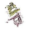
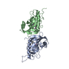
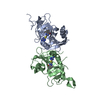
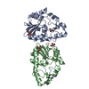
- PDB-3t61: Crystal Structure of a gluconokinase from Sinorhizobium meliloti 1021 -
+ Open data

- Basic information
| Entry | Database: PDB / ID: 3t61 | ||||||
|---|---|---|---|---|---|---|---|
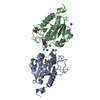
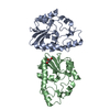
| Title | Crystal Structure of a gluconokinase from Sinorhizobium meliloti 1021 | ||||||
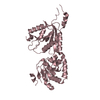
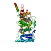
 Components Components | Gluconokinase | ||||||
 Keywords Keywords | TRANSFERASE / PSI-BIOLOGY / Structural Genomics / Protein Structure Initiative / New York Structural Genomics Research Consortium / NYSGRC / Alpha/Beta / gluconokinase / Kinase / Gluconic acid | ||||||
| Function / homology |  Function and homology information Function and homology informationgluconokinase / gluconokinase activity / D-gluconate metabolic process / ATP binding / cytoplasm Similarity search - Function | ||||||
| Biological species |  Sinorhizobium meliloti (bacteria) Sinorhizobium meliloti (bacteria) | ||||||
| Method |  X-RAY DIFFRACTION / SYNCHROTRON / SAD / Resolution: 2.2 Å X-RAY DIFFRACTION / SYNCHROTRON / SAD / Resolution: 2.2 Å | ||||||
 Authors Authors | Kumaran, D. / Chamala, S. / Evans, B. / Foti, R. / Gizzi, A. / Hillerich, B. / Kar, A. / LaFleur, J. / Seidel, R. / Villigas, G. ...Kumaran, D. / Chamala, S. / Evans, B. / Foti, R. / Gizzi, A. / Hillerich, B. / Kar, A. / LaFleur, J. / Seidel, R. / Villigas, G. / Zencheck, W. / Almo, S.C. / Swaminathan, S. / New York Structural Genomics Research Consortium (NYSGRC) | ||||||
 Citation Citation | Journal: To be Published Title: Crystal Structure of a gluconokinase from Sinorhizobium meliloti 1021 Authors: Kumaran, D. / Almo, S.C. / Swaminathan, S. | ||||||
| History |
|
- Structure visualization
| Structure viewer | Molecule: MolmilJmol/JSmol |
|---|
- Downloads & links
-Download
| PDBx/mmCIF format | 3t61.cif.gz | 84 KB | Display | PDBx/mmCIF format |
|---|---|---|---|---|
| PDB format | pdb3t61.ent.gz | 63.3 KB | Display | PDB format |
| PDBx/mmJSON format | 3t61.json.gz | Tree view | PDBx/mmJSON format | |
| Others |  Other downloads Other downloads |
-Validation report
| Arichive directory | https://data.pdbj.org/pub/pdb/validation_reports/t6/3t61ftp://data.pdbj.org/pub/pdb/validation_reports/t6/3t61 | HTTPS FTP |
|---|
-Related structure data
| Similar structure data | |
|---|---|
| Other databases |


-Links
 PDBj
PDBj- Assembly
| Deposited unit | 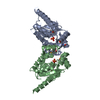
| ||||||||
|---|---|---|---|---|---|---|---|---|---|
| 1 |
| ||||||||
| Unit cell |
| ||||||||
| Details | Dimer |
-Components
| #1: Protein | Mass: 22654.764 Da / Num. of mol.: 2 Source method: isolated from a genetically manipulated source Source: (gene. exp.) Sinorhizobium meliloti (bacteria) / Strain: 1021 / Gene: gntK, RB0698, SM_b21119 / Plasmid: pET3A / Production host: #2: Chemical |   Mass: 94.971 Da / Num. of mol.: 2 / Source method: obtained synthetically / Formula: PO4 Mass: 94.971 Da / Num. of mol.: 2 / Source method: obtained synthetically / Formula: PO4#3: Water | ChemComp-HOH / |  Mass: 18.015 Da / Num. of mol.: 154 / Source method: isolated from a natural source / Formula: H2O Mass: 18.015 Da / Num. of mol.: 154 / Source method: isolated from a natural source / Formula: H2OHas protein modification | Y | |
|---|
-Experimental details
-Experiment
| Experiment | Method: X-RAY DIFFRACTION / Number of used crystals: 1 |
|---|
- Sample preparation
| Crystal | Density Matthews: 3.04 Å3/Da / Density % sol: 59.6 % |
|---|---|
| Crystal grow | Temperature: 293 K / Method: vapor diffusion, sitting drop / pH: 8.5 Details: 2M Ammonium Phosphate Monobasic, 0.1 M Tris, 2% sucrose, pH 8.5, VAPOR DIFFUSION, SITTING DROP, temperature 293K |
-Data collection
| Diffraction | Mean temperature: 100 K |
|---|---|
| Diffraction source | Source: SYNCHROTRON / Site: NSLS  / Beamline: X29A / Wavelength: 0.9795 Å / Beamline: X29A / Wavelength: 0.9795 Å |
| Detector | Type: ADSC QUANTUM 315 / Detector: CCD / Date: Jul 27, 2011 / Details: mirrors |
| Radiation | Monochromator: Si III / Protocol: SINGLE WAVELENGTH / Monochromatic (M) / Laue (L): M / Scattering type: x-ray |
| Radiation wavelength | Wavelength: 0.9795 Å / Relative weight: 1 |
| Reflection | Resolution: 2.2→50 Å / Num. all: 27740 / Num. obs: 27740 / % possible obs: 100 % / Observed criterion σ(F): 0 / Observed criterion σ(I): 0 / Redundancy: 17.1 % / Biso Wilson estimate: 24.7 Å2 / Rmerge(I) obs: 0.097 / Net I/σ(I): 16.5 |
| Reflection shell | Resolution: 2.2→2.26 Å / Redundancy: 16.9 % / Rmerge(I) obs: 0.506 / Mean I/σ(I) obs: 2.5 / Num. unique all: 2284 / % possible all: 100 |
- Processing
| Software |
| |||||||||||||||||||||||||
|---|---|---|---|---|---|---|---|---|---|---|---|---|---|---|---|---|---|---|---|---|---|---|---|---|---|---|
| Refinement | Method to determine structure: SAD / Resolution: 2.2→44.68 Å / Rfactor Rfree error: 0.006 / Data cutoff high absF: 169839.34 / Data cutoff low absF: 0 / Isotropic thermal model: RESTRAINED / Cross valid method: THROUGHOUT / σ(F): 0 / Stereochemistry target values: Engh & Huber Details: Extra density near the phosphate ion is not modeled.
| |||||||||||||||||||||||||
| Solvent computation | Solvent model: FLAT MODEL / Bsol: 39.2911 Å2 / ksol: 0.343718 e/Å3 | |||||||||||||||||||||||||
| Displacement parameters | Biso mean: 37.2 Å2
| |||||||||||||||||||||||||
| Refine analyze |
| |||||||||||||||||||||||||
| Refinement step | Cycle: LAST / Resolution: 2.2→44.68 Å
| |||||||||||||||||||||||||
| Refine LS restraints |
| |||||||||||||||||||||||||
| Refine LS restraints NCS | NCS model details: NONE | |||||||||||||||||||||||||
| LS refinement shell | Resolution: 2.2→2.34 Å / Rfactor Rfree error: 0.019 / Total num. of bins used: 6
| |||||||||||||||||||||||||
| Xplor file |
|
