Resolution: 2.86→3.04 Å / Redundancy: 8.5 % / Rmerge(I) obs: 0.5 / Mean I/σ(I) obs: 1.5 / Rsym value: 0.5 / % possible all: 60.5
-
Processing
Software
Name
Version
Classification
Blu-Ice
datacollection
SHARP
phasing
PHENIX
(phenix.refine: 1.6.4_486)
refinement
MOSFLM
datareduction
SCALA
datascaling
Refinement
Method to determine structure: MIR / Resolution: 2.869→43.057 Å / SU ML: 0.42 / σ(F): 1.34 / Phase error: 27.34 / Stereochemistry target values: ML Details: THE LAST DOMAIN (RESIDUES 834-913) WAS BUILT BY THREADING THE AMINO ACID SEQUENCE ON THE NMR STRUCTURE OF SAME DOMAIN FROM C7 (PDB ENTRY 2WCY). SINCE THE ELECTRON DENSITY WAS WEAK, THE ...Details: THE LAST DOMAIN (RESIDUES 834-913) WAS BUILT BY THREADING THE AMINO ACID SEQUENCE ON THE NMR STRUCTURE OF SAME DOMAIN FROM C7 (PDB ENTRY 2WCY). SINCE THE ELECTRON DENSITY WAS WEAK, THE SEQUENCE ANNOTATION IS NOT RELIABLE.
Rfactor
Num. reflection
% reflection
Rfree
0.2759
2179
6.1 %
Rwork
0.2217
-
-
obs
0.225
35743
94.92 %
Solvent computation
Shrinkage radii: 0.72 Å / VDW probe radii: 1 Å / Solvent model: FLAT BULK SOLVENT MODEL / Bsol: 69.704 Å2 / ksol: 0.313 e/Å3
In the structure databanks used in Yorodumi, some data are registered as the other names, "COVID-19 virus" and "2019-nCoV". Here are the details of the virus and the list of structure data.
Jan 31, 2019. EMDB accession codes are about to change! (news from PDBe EMDB page)
EMDB accession codes are about to change! (news from PDBe EMDB page)
The allocation of 4 digits for EMDB accession codes will soon come to an end. Whilst these codes will remain in use, new EMDB accession codes will include an additional digit and will expand incrementally as the available range of codes is exhausted. The current 4-digit format prefixed with “EMD-” (i.e. EMD-XXXX) will advance to a 5-digit format (i.e. EMD-XXXXX), and so on. It is currently estimated that the 4-digit codes will be depleted around Spring 2019, at which point the 5-digit format will come into force.
The EM Navigator/Yorodumi systems omit the EMD- prefix.
Related info.:Q: What is EMD? / ID/Accession-code notation in Yorodumi/EM Navigator
Yorodumi is a browser for structure data from EMDB, PDB, SASBDB, etc.
This page is also the successor to EM Navigator detail page, and also detail information page/front-end page for Omokage search.
The word "yorodu" (or yorozu) is an old Japanese word meaning "ten thousand". "mi" (miru) is to see.
Related info.:EMDB / PDB / SASBDB / Comparison of 3 databanks / Yorodumi Search / Aug 31, 2016. New EM Navigator & Yorodumi / Yorodumi Papers / Jmol/JSmol / Function and homology information / Changes in new EM Navigator and Yorodumi
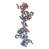
 Movie
Movie Controller


 Open data
 Basic information
 Components
 Keywords
 Function and homology information
 Homo sapiens (human)
 X-RAY DIFFRACTION /
  Authors
 Citation
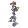
 Structure visualization
 Downloads & links
 Other downloads
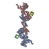

 PDBj
PDBj


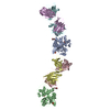
 Assembly

 Mass: 112.411 Da / Num. of mol.: 1 / Source method: obtained synthetically / Formula: Cd
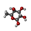

 Type: L-saccharide, beta linking / Mass: 164.156 Da / Num. of mol.: 1
 Type: D-saccharide, alpha linking / Mass: 180.156 Da / Num. of mol.: 4
 Sample preparation
 / Beamline: BL9-2 / Wavelength: 0.991
  Processing