 Movie
Movie Controller

[English] 日本語
 Yorodumi
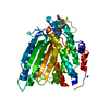
- PDB-3t5a: Crystal structure of N-terminal domain of FAAL28 G330W mutant fro... -
+ Open data

- Basic information
| Entry | Database: PDB / ID: 3t5a | ||||||
|---|---|---|---|---|---|---|---|
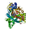
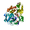
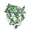
| Title | Crystal structure of N-terminal domain of FAAL28 G330W mutant from Mycobacterium tuberculosis | ||||||
 Components Components | Long-chain-fatty-acid--AMP ligase FadD28 | ||||||
 Keywords Keywords | LIGASE / Acetyl-CoA synthetase like fold / AMP-binding | ||||||
| Function / homology |  Function and homology information Function and homology informationlong-chain fatty acid adenylyltransferase FadD28 / Dimycocersyl phthiocerol biosynthesis / adenylyltransferase activity / fatty-acyl-CoA synthase activity / response to host immune response / DIM/DIP cell wall layer assembly / biological process involved in interaction with host / lipid biosynthetic process / ligase activity / fatty acid biosynthetic process ...long-chain fatty acid adenylyltransferase FadD28 / Dimycocersyl phthiocerol biosynthesis / adenylyltransferase activity / fatty-acyl-CoA synthase activity / response to host immune response / DIM/DIP cell wall layer assembly / biological process involved in interaction with host / lipid biosynthetic process / ligase activity / fatty acid biosynthetic process / symbiont-mediated suppression of host innate immune response / ATP binding / plasma membrane / cytosol Similarity search - Function | ||||||
| Biological species |   Mycobacterium tuberculosis (bacteria) Mycobacterium tuberculosis (bacteria) | ||||||
| Method |  X-RAY DIFFRACTION / MOLECULAR REPLACEMENT / Resolution: 2.05 Å X-RAY DIFFRACTION / MOLECULAR REPLACEMENT / Resolution: 2.05 Å | ||||||
 Authors Authors | Goyal, A. / Sankaranarayanan, R. | ||||||
 Citation Citation | Journal: J.Mol.Biol. / Year: 2012 Title: Molecular basis of the functional divergence of fatty acyl-AMP ligase biosynthetic enzymes of Mycobacterium tuberculosis. Authors: Goyal, A. / Verma, P. / Anandhakrishnan, M. / Gokhale, R.S. / Sankaranarayanan, R. | ||||||
| History |
|
- Structure visualization
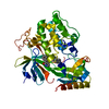
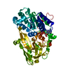
| Structure viewer | Molecule: MolmilJmol/JSmol |
|---|
- Downloads & links
-Download
| PDBx/mmCIF format | 3t5a.cif.gz | 104.6 KB | Display | PDBx/mmCIF format |
|---|---|---|---|---|
| PDB format | pdb3t5a.ent.gz | 77.7 KB | Display | PDB format |
| PDBx/mmJSON format | 3t5a.json.gz | Tree view | PDBx/mmJSON format | |
| Others |  Other downloads Other downloads |
-Validation report
| Summary document | 3t5a_validation.pdf.gz | 428.6 KB | Display | wwPDB validaton report |
|---|---|---|---|---|
| Full document | 3t5a_full_validation.pdf.gz | 440.6 KB | Display | |
| Data in XML | 3t5a_validation.xml.gz | 21.6 KB | Display | |
| Data in CIF | 3t5a_validation.cif.gz | 31.7 KB | Display | |
| Arichive directory | https://data.pdbj.org/pub/pdb/validation_reports/t5/3t5aftp://data.pdbj.org/pub/pdb/validation_reports/t5/3t5a | HTTPS FTP |
-Related structure data
| Related structure data | 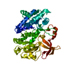 3t5bC 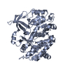 3t5cC 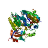 3e53S S: Starting model for refinement C: citing same article ( |
|---|---|
| Similar structure data |


-Links
 PDBj
PDBj
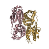
- Assembly
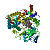
| Deposited unit | 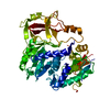
| ||||||||
|---|---|---|---|---|---|---|---|---|---|
| 1 |
| ||||||||
| Unit cell |
|
-Components
| #1: Protein | Mass: 52073.285 Da / Num. of mol.: 1 / Fragment: N-terminal domain (UNP RESIDUES 1-460) / Mutation: G330W Source method: isolated from a genetically manipulated source Source: (gene. exp.) Mycobacterium tuberculosis (bacteria) / Strain: H37Rv / Gene: acoas, fadD28, MT3011, Rv2941 / Plasmid: pET28c / Production host: References: UniProt: P96290, UniProt: P9WQ59*PLUS, Ligases; Forming carbon-sulfur bonds; Acid-thiol ligases |
|---|---|
| #2: Water | ChemComp-HOH /  Mass: 18.015 Da / Num. of mol.: 323 / Source method: isolated from a natural source / Formula: H2O Mass: 18.015 Da / Num. of mol.: 323 / Source method: isolated from a natural source / Formula: H2O |
-Experimental details
-Experiment
| Experiment | Method: X-RAY DIFFRACTION / Number of used crystals: 1 |
|---|
- Sample preparation
| Crystal | Density Matthews: 2.04 Å3/Da / Density % sol: 39.79 % |
|---|---|
| Crystal grow | Temperature: 277 K / Method: vapor diffusion, hanging drop / pH: 6.3 Details: 18% PEG 3350, 0.1M Sodium MES, 5% ethylene glycol, 0.15M Lithium sulphate, pH 6.3, VAPOR DIFFUSION, HANGING DROP, temperature 277K |
-Data collection
| Diffraction | Mean temperature: 100 K |
|---|---|
| Diffraction source | Source: ROTATING ANODE / Type: RIGAKU RUH3R / Wavelength: 1.5418 Å |
| Detector | Type: MAR scanner 345 mm plate / Detector: IMAGE PLATE / Date: Nov 28, 2008 / Details: Osmic mirrors |
| Radiation | Monochromator: Ni FILTER / Protocol: SINGLE WAVELENGTH / Monochromatic (M) / Laue (L): M / Scattering type: x-ray |
| Radiation wavelength | Wavelength: 1.5418 Å / Relative weight: 1 |
| Reflection | Resolution: 2.05→25 Å / Num. obs: 25902 / % possible obs: 92.9 % / Observed criterion σ(I): 2 / Redundancy: 4.7 % / Biso Wilson estimate: 31.69 Å2 / Rmerge(I) obs: 0.098 / Net I/σ(I): 14.63 |
| Reflection shell | Resolution: 2.05→2.12 Å / Redundancy: 4.4 % / Rmerge(I) obs: 0.486 / Mean I/σ(I) obs: 2.03 / Num. unique all: 2564 / % possible all: 94 |
- Processing
| Software |
| ||||||||||||||||||||
|---|---|---|---|---|---|---|---|---|---|---|---|---|---|---|---|---|---|---|---|---|---|
| Refinement | Method to determine structure: MOLECULAR REPLACEMENT Starting model: PDB ENTRY 3E53 Resolution: 2.05→25 Å / σ(F): 2
| ||||||||||||||||||||
| Displacement parameters |
| ||||||||||||||||||||
| Refinement step | Cycle: LAST / Resolution: 2.05→25 Å
|
