 Movie
Movie Controller

[English] 日本語
 Yorodumi
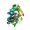
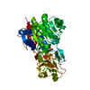
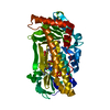
- PDB-3t00: Crystal Structure of Phosphonoacetate hydrolase from Sinorhizobiu... -
+ Open data

- Basic information
| Entry | Database: PDB / ID: 3t00 | ||||||
|---|---|---|---|---|---|---|---|
| Title | Crystal Structure of Phosphonoacetate hydrolase from Sinorhizobium meliloti 1021 in complex with vanadate | ||||||
 Components Components | phosphonoacetate hydrolase | ||||||
 Keywords Keywords | HYDROLASE / alkaline phosphatase superfamily / transition state mimic | ||||||
| Function / homology |  Function and homology information Function and homology informationphosphonoacetate hydrolase activity / nucleotide diphosphatase / nucleoside triphosphate diphosphatase activity / metal ion binding Similarity search - Function | ||||||
| Biological species |  Sinorhizobium meliloti (bacteria) Sinorhizobium meliloti (bacteria) | ||||||
| Method |  X-RAY DIFFRACTION / SYNCHROTRON / MOLECULAR REPLACEMENT / Resolution: 1.8 Å X-RAY DIFFRACTION / SYNCHROTRON / MOLECULAR REPLACEMENT / Resolution: 1.8 Å | ||||||
 Authors Authors | Agarwal, V. / Nair, S.K. | ||||||
 Citation Citation | Journal: Chem.Biol. / Year: 2011 Title: Structural and mechanistic insights into C-p bond hydrolysis by phosphonoacetate hydrolase. Authors: Agarwal, V. / Borisova, S.A. / Metcalf, W.W. / van der Donk, W.A. / Nair, S.K. | ||||||
| History |
|
- Structure visualization
| Structure viewer | Molecule: MolmilJmol/JSmol |
|---|
- Downloads & links
-Download
| PDBx/mmCIF format | 3t00.cif.gz | 103.3 KB | Display | PDBx/mmCIF format |
|---|---|---|---|---|
| PDB format | pdb3t00.ent.gz | 76.5 KB | Display | PDB format |
| PDBx/mmJSON format | 3t00.json.gz | Tree view | PDBx/mmJSON format | |
| Others |  Other downloads Other downloads |
-Validation report
| Arichive directory | https://data.pdbj.org/pub/pdb/validation_reports/t0/3t00ftp://data.pdbj.org/pub/pdb/validation_reports/t0/3t00 | HTTPS FTP |
|---|
-Related structure data
| Related structure data |  3szySC  3szzC  3t01C  3t02C S: Starting model for refinement C: citing same article ( |
|---|---|
| Similar structure data |


-Links
 PDBj
PDBj
- Assembly
| Deposited unit | 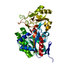
| ||||||||
|---|---|---|---|---|---|---|---|---|---|
| 1 |
| ||||||||
| Unit cell |
|
-Components
| #1: Protein | Mass: 46593.875 Da / Num. of mol.: 1 Source method: isolated from a genetically manipulated source Source: (gene. exp.) Sinorhizobium meliloti (bacteria) / Strain: 1021 / Gene: phnA, RB0978, SM_b21538 / Plasmid: pET28 / Production host: | ||||||
|---|---|---|---|---|---|---|---|
| #2: Chemical |   Mass: 65.409 Da / Num. of mol.: 2 / Source method: obtained synthetically / Formula: Zn Mass: 65.409 Da / Num. of mol.: 2 / Source method: obtained synthetically / Formula: Zn#3: Chemical | ChemComp-VO4 / |   Mass: 114.939 Da / Num. of mol.: 1 / Source method: obtained synthetically / Formula: VO4 Mass: 114.939 Da / Num. of mol.: 1 / Source method: obtained synthetically / Formula: VO4#4: Chemical | ChemComp-NI / |   Mass: 58.693 Da / Num. of mol.: 1 / Source method: obtained synthetically / Formula: Ni Mass: 58.693 Da / Num. of mol.: 1 / Source method: obtained synthetically / Formula: Ni#5: Water | ChemComp-HOH / |  Mass: 18.015 Da / Num. of mol.: 351 / Source method: isolated from a natural source / Formula: H2O Mass: 18.015 Da / Num. of mol.: 351 / Source method: isolated from a natural source / Formula: H2O |
-Experimental details
-Experiment
| Experiment | Method: X-RAY DIFFRACTION / Number of used crystals: 1 |
|---|
- Sample preparation
| Crystal | Density Matthews: 2.4 Å3/Da / Density % sol: 48.74 % |
|---|---|
| Crystal grow | Temperature: 298 K / Method: vapor diffusion, hanging drop / pH: 7.5 Details: 20% PEG3350, 0.2 M NaCl, 0.1 M HEPES, pH 7.5, VAPOR DIFFUSION, HANGING DROP, temperature 298K |
-Data collection
| Diffraction | Mean temperature: 100 K |
|---|---|
| Diffraction source | Source: SYNCHROTRON / Site: APS  / Beamline: 21-ID-D / Wavelength: 0.97857 / Beamline: 21-ID-D / Wavelength: 0.97857 |
| Detector | Type: MARMOSAIC 300 mm CCD / Detector: CCD / Date: Dec 20, 2010 |
| Radiation | Monochromator: KOHZU / Protocol: SINGLE WAVELENGTH / Monochromatic (M) / Laue (L): M / Scattering type: x-ray |
| Radiation wavelength | Wavelength: 0.97857 Å / Relative weight: 1 |
| Reflection | Resolution: 1.8→50 Å / Num. all: 42647 / Num. obs: 41825 / % possible obs: 100 % / Redundancy: 13.8 % / Rmerge(I) obs: 0.053 / Χ2: 1.335 / Net I/σ(I): 13.7 |
| Reflection shell | Resolution: 1.8→1.83 Å / Rmerge(I) obs: 0.693 / Mean I/σ(I) obs: 12.1 |
- Processing
| Software |
| |||||||||||||||||||||||||||||||||||||||||||||||||||||||||||||||||
|---|---|---|---|---|---|---|---|---|---|---|---|---|---|---|---|---|---|---|---|---|---|---|---|---|---|---|---|---|---|---|---|---|---|---|---|---|---|---|---|---|---|---|---|---|---|---|---|---|---|---|---|---|---|---|---|---|---|---|---|---|---|---|---|---|---|---|
| Refinement | Method to determine structure: MOLECULAR REPLACEMENT Starting model: PDB ENTRY 3SZY Resolution: 1.8→25 Å / Cor.coef. Fo:Fc: 0.947 / Cor.coef. Fo:Fc free: 0.921 / Occupancy max: 1 / Occupancy min: 0.5 / SU B: 2.57 / SU ML: 0.082 / Cross valid method: THROUGHOUT / σ(F): 0 / ESU R: 0.141 / ESU R Free: 0.133 / Stereochemistry target values: MAXIMUM LIKELIHOOD Details: HYDROGENS HAVE BEEN ADDED IN THE RIDING POSITIONS U VALUES : REFINED INDIVIDUALLY
| |||||||||||||||||||||||||||||||||||||||||||||||||||||||||||||||||
| Solvent computation | Ion probe radii: 0.8 Å / Shrinkage radii: 0.8 Å / VDW probe radii: 1.4 Å / Solvent model: BABINET MODEL WITH MASK | |||||||||||||||||||||||||||||||||||||||||||||||||||||||||||||||||
| Displacement parameters | Biso max: 67.47 Å2 / Biso mean: 28.9543 Å2 / Biso min: 10.68 Å2
| |||||||||||||||||||||||||||||||||||||||||||||||||||||||||||||||||
| Refinement step | Cycle: LAST / Resolution: 1.8→25 Å
| |||||||||||||||||||||||||||||||||||||||||||||||||||||||||||||||||
| Refine LS restraints |
| |||||||||||||||||||||||||||||||||||||||||||||||||||||||||||||||||
| LS refinement shell | Resolution: 1.8→1.847 Å / Total num. of bins used: 20
|
