 Movie
Movie Controller

[English] 日本語
 Yorodumi
- PDB-1nt4: Crystal structure of Escherichia coli periplasmic glucose-1-phosp... -
+ Open data

- Basic information
| Entry | Database: PDB / ID: 1nt4 | |||||||||
|---|---|---|---|---|---|---|---|---|---|---|
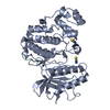
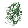
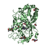
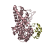
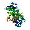
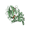
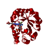
| Title | Crystal structure of Escherichia coli periplasmic glucose-1-phosphatase H18A mutant complexed with glucose-1-phosphate | |||||||||
 Components Components | Glucose-1-phosphatase | |||||||||
 Keywords Keywords | HYDROLASE / alpha domain / alpha-beta domain / occluded active site / enzyme-substrate complex / Montreal-Kingston Bacterial Structural Genomics Initiative / BSGI / Structural Genomics | |||||||||
| Function / homology |  Function and homology information Function and homology informationglucose-1-phosphatase / inositol hexakisphosphate 3-phosphatase activity / glucose-1-phosphatase activity / sugar-phosphatase activity / glucose catabolic process / outer membrane-bounded periplasmic space Similarity search - Function | |||||||||
| Biological species |  | |||||||||
| Method |  X-RAY DIFFRACTION / SYNCHROTRON / MAD / Resolution: 2.4 Å X-RAY DIFFRACTION / SYNCHROTRON / MAD / Resolution: 2.4 Å | |||||||||
 Authors Authors | Lee, D.C. / Cottrill, M.A. / Forsberg, C.W. / Jia, Z. / Montreal-Kingston Bacterial Structural Genomics Initiative (BSGI) | |||||||||
 Citation Citation | Journal: J.Biol.Chem. / Year: 2003 Title: Functional insights revealed by the crystal structures of Escherichia coli glucose-1-phosphatase. Authors: Lee, D.C. / Cottrill, M.A. / Forsberg, C.W. / Jia, Z. | |||||||||
| History |
|
- Structure visualization
| Structure viewer | Molecule: MolmilJmol/JSmol |
|---|
- Downloads & links
-Download
| PDBx/mmCIF format | 1nt4.cif.gz | 170.4 KB | Display | PDBx/mmCIF format |
|---|---|---|---|---|
| PDB format | pdb1nt4.ent.gz | 134.8 KB | Display | PDB format |
| PDBx/mmJSON format | 1nt4.json.gz | Tree view | PDBx/mmJSON format | |
| Others |  Other downloads Other downloads |
-Validation report
| Arichive directory | https://data.pdbj.org/pub/pdb/validation_reports/nt/1nt4ftp://data.pdbj.org/pub/pdb/validation_reports/nt/1nt4 | HTTPS FTP |
|---|
-Related structure data
| Similar structure data | |
|---|---|
| Other databases |


-Links
 PDBj
PDBj


- Assembly
| Deposited unit | 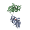
| ||||||||
|---|---|---|---|---|---|---|---|---|---|
| 1 | 
| ||||||||
| 2 | 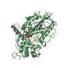
| ||||||||
| Unit cell |
|
-Components
| #1: Protein | Mass: 43537.117 Da / Num. of mol.: 2 / Mutation: H18A Source method: isolated from a genetically manipulated source Source: (gene. exp.) #2: Sugar |   Type: D-saccharide / Mass: 260.136 Da / Num. of mol.: 2 Type: D-saccharide / Mass: 260.136 Da / Num. of mol.: 2Source method: isolated from a genetically manipulated source Formula: C6H13O9P #3: Water | ChemComp-HOH / |  Mass: 18.015 Da / Num. of mol.: 351 / Source method: isolated from a natural source / Formula: H2O Mass: 18.015 Da / Num. of mol.: 351 / Source method: isolated from a natural source / Formula: H2OHas protein modification | Y | |
|---|
-Experimental details
-Experiment
| Experiment | Method: X-RAY DIFFRACTION / Number of used crystals: 1 |
|---|
- Sample preparation
| Crystal | Density Matthews: 2.28 Å3/Da / Density % sol: 46.07 % | ||||||||||||||||||||||||||||||
|---|---|---|---|---|---|---|---|---|---|---|---|---|---|---|---|---|---|---|---|---|---|---|---|---|---|---|---|---|---|---|---|
| Crystal grow | Temperature: 293 K / Method: vapor diffusion, hanging drop / pH: 6.5 Details: Ammonium sulfate, PEG 500 mme, pH 6.5, VAPOR DIFFUSION, HANGING DROP, temperature 293K | ||||||||||||||||||||||||||||||
| Crystal grow | *PLUS pH: 6.25 / Method: vapor diffusion, hanging drop | ||||||||||||||||||||||||||||||
| Components of the solutions | *PLUS
|
-Data collection
| Diffraction | Mean temperature: 100 K | ||||||||||||
|---|---|---|---|---|---|---|---|---|---|---|---|---|---|
| Diffraction source | Source: SYNCHROTRON / Site: CHESS  / Beamline: F1 / Wavelength: 1.2146, 1.2149, 1.3190 / Beamline: F1 / Wavelength: 1.2146, 1.2149, 1.3190 | ||||||||||||
| Detector | Type: ADSC QUANTUM 4 / Detector: CCD / Date: Feb 7, 2001 | ||||||||||||
| Radiation | Protocol: MAD / Monochromatic (M) / Laue (L): M / Scattering type: x-ray | ||||||||||||
| Radiation wavelength |
| ||||||||||||
| Reflection | Resolution: 2.4→30 Å / Num. all: 27106 / Num. obs: 27106 / % possible obs: 91.9 % / Observed criterion σ(F): 1 / Observed criterion σ(I): 1 / Redundancy: 3.45 % / Rsym value: 0.078 / Net I/σ(I): 17.4 | ||||||||||||
| Reflection shell | Resolution: 2.4→2.5 Å / Redundancy: 2.3 % / Mean I/σ(I) obs: 3.5 / Num. unique all: 3023 / Rsym value: 0.271 / % possible all: 91.1 | ||||||||||||
| Reflection | *PLUS Num. obs: 27760 / Num. measured all: 95761 / Rmerge(I) obs: 0.078 | ||||||||||||
| Reflection shell | *PLUS % possible obs: 91.1 % / Rmerge(I) obs: 0.271 |
- Processing
| Software |
| |||||||||||||||||||||||||
|---|---|---|---|---|---|---|---|---|---|---|---|---|---|---|---|---|---|---|---|---|---|---|---|---|---|---|
| Refinement | Method to determine structure: MAD / Resolution: 2.4→30 Å / Isotropic thermal model: isotropic / Cross valid method: THROUGHOUT / σ(F): 1 / Stereochemistry target values: Engh & Huber
| |||||||||||||||||||||||||
| Displacement parameters | Biso mean: 29.75 Å2 | |||||||||||||||||||||||||
| Refinement step | Cycle: LAST / Resolution: 2.4→30 Å
| |||||||||||||||||||||||||
| Refine LS restraints |
| |||||||||||||||||||||||||
| LS refinement shell | Resolution: 2.4→2.49 Å
| |||||||||||||||||||||||||
| Refinement | *PLUS % reflection Rfree: 5 % / Rfactor Rfree: 0.284 / Rfactor Rwork: 0.2179 | |||||||||||||||||||||||||
| Solvent computation | *PLUS | |||||||||||||||||||||||||
| Displacement parameters | *PLUS |
