 Movie
Movie Controller

[English] 日本語
 Yorodumi
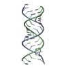
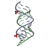
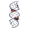
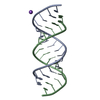
- PDB-3syw: Crystal Structure of the Triplet Repeat in Myotonic Dystrophy Rev... -
+ Open data

- Basic information
| Entry | Database: PDB / ID: 3syw | ||||||||||||||||||
|---|---|---|---|---|---|---|---|---|---|---|---|---|---|---|---|---|---|---|---|
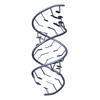
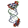
| Title | Crystal Structure of the Triplet Repeat in Myotonic Dystrophy Reveals Heterogeneous 1x1 Nucleotide UU Internal Loop Conformations | ||||||||||||||||||
 Components Components | RNA (5'-R(* Keywords KeywordsRNA / CUG repeat RNA / A-form RNA / Myotonic dystrophy / Internal loop | Function / homology | PHOSPHATE ION / RNA / RNA (> 10) |  Function and homology information Function and homology informationMethod |  X-RAY DIFFRACTION / SYNCHROTRON / MOLECULAR REPLACEMENT / Resolution: 1.57 Å X-RAY DIFFRACTION / SYNCHROTRON / MOLECULAR REPLACEMENT / Resolution: 1.57 Å  Authors AuthorsKumar, A. / Park, H. / Pengfei, F. / Parkesh, R. / Guo, M. / Nettles, K.W. / Disney, M.D. |  CitationJournal: Biochemistry / Year: 2011 CitationJournal: Biochemistry / Year: 2011Title: Crystal Structure of the Triplet Repeat in Myotonic Dystrophy Reveals Heterogeneous 1x1 Nucleotide UU Internal Loop Conformations Authors: Kumar, A. / Park, H. / Fang, P. / Parkesh, R. / Guo, M. / Nettles, K.W. / Disney, M.D. History |
|
- Structure visualization
| Structure viewer | Molecule: MolmilJmol/JSmol |
|---|
- Downloads & links
-Download
| PDBx/mmCIF format | 3syw.cif.gz | 66 KB | Display | PDBx/mmCIF format |
|---|---|---|---|---|
| PDB format | pdb3syw.ent.gz | 52 KB | Display | PDB format |
| PDBx/mmJSON format | 3syw.json.gz | Tree view | PDBx/mmJSON format | |
| Others |  Other downloads Other downloads |
-Validation report
| Summary document | 3syw_validation.pdf.gz | 419.8 KB | Display | wwPDB validaton report |
|---|---|---|---|---|
| Full document | 3syw_full_validation.pdf.gz | 421.7 KB | Display | |
| Data in XML | 3syw_validation.xml.gz | 6.3 KB | Display | |
| Data in CIF | 3syw_validation.cif.gz | 8.8 KB | Display | |
| Arichive directory | https://data.pdbj.org/pub/pdb/validation_reports/sy/3sywftp://data.pdbj.org/pub/pdb/validation_reports/sy/3syw | HTTPS FTP |
-Related structure data

-Links
 PDBj
PDBj


- Assembly
| Deposited unit | 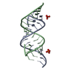
| |||||||||||||||||||||||||||||||||
|---|---|---|---|---|---|---|---|---|---|---|---|---|---|---|---|---|---|---|---|---|---|---|---|---|---|---|---|---|---|---|---|---|---|---|
| 1 |
| |||||||||||||||||||||||||||||||||
| 2 | 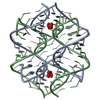
| |||||||||||||||||||||||||||||||||
| Unit cell |
| |||||||||||||||||||||||||||||||||
| Components on special symmetry positions |
|
-Components
| #1: RNA chain | Mass: 6039.569 Da / Num. of mol.: 2 / Fragment: RNA / Source method: obtained synthetically / Details: Polyribonucleotide #2: Chemical |   Mass: 94.971 Da / Num. of mol.: 2 / Source method: obtained synthetically / Formula: PO4 / Details: Polyribonucleotide Mass: 94.971 Da / Num. of mol.: 2 / Source method: obtained synthetically / Formula: PO4 / Details: Polyribonucleotide#3: Water | ChemComp-HOH / |  Mass: 18.015 Da / Num. of mol.: 176 / Source method: isolated from a natural source / Formula: H2O Mass: 18.015 Da / Num. of mol.: 176 / Source method: isolated from a natural source / Formula: H2O |
|---|
-Experimental details
-Experiment
| Experiment | Method: X-RAY DIFFRACTION / Number of used crystals: 2 |
|---|
- Sample preparation
| Crystal | Density Matthews: 2.33 Å3/Da / Density % sol: 47.18 % |
|---|---|
| Crystal grow | Temperature: 291 K / pH: 6 Details: 15 mM magnesium acetate, 50 mM sodium cacodylate, pH 6.0, 1.7 M ammonium sulfate, VAPOR DIFFUSION, SITTING DROP, temperature 291K |
-Data collection
| Diffraction | Mean temperature: 100 K |
|---|---|
| Diffraction source | Source: SYNCHROTRON / Site: SSRL  / Beamline: BL9-2 / Wavelength: 0.97855 / Beamline: BL9-2 / Wavelength: 0.97855 |
| Detector | Type: MARMOSAIC 325 mm CCD / Detector: CCD / Date: Jan 9, 2011 / Details: RH COATED FLAT MIRROR, TOROIDAL FOCUSING MIRROR |
| Radiation | Monochromator: DOUBLE CRYSTAL MONOCHROMATOR / Protocol: SINGLE WAVELENGTH / Monochromatic (M) / Laue (L): M / Scattering type: x-ray |
| Radiation wavelength | Wavelength: 0.97855 Å / Relative weight: 1 |
| Reflection | Resolution: 1.52→34.6 Å / Num. obs: 16712 / % possible obs: 99.5 % / Observed criterion σ(I): 1 / Rmerge(I) obs: 0.057 / Net I/σ(I): 48.99 |
| Reflection shell | Resolution: 1.52→1.57 Å / Redundancy: 4.2 % / Rmerge(I) obs: 0.487 / Mean I/σ(I) obs: 3.1 / % possible all: 98.9 |
- Processing
| Software |
| |||||||||||||||||||||||||||||||||||||||||||||||||
|---|---|---|---|---|---|---|---|---|---|---|---|---|---|---|---|---|---|---|---|---|---|---|---|---|---|---|---|---|---|---|---|---|---|---|---|---|---|---|---|---|---|---|---|---|---|---|---|---|---|---|
| Refinement | Method to determine structure: MOLECULAR REPLACEMENT / Resolution: 1.57→34.6 Å / σ(F): 0.15
| |||||||||||||||||||||||||||||||||||||||||||||||||
| Refinement step | Cycle: LAST / Resolution: 1.57→34.6 Å
| |||||||||||||||||||||||||||||||||||||||||||||||||
| Refine LS restraints |
| |||||||||||||||||||||||||||||||||||||||||||||||||
| LS refinement shell |
|
