 Movie
Movie Controller

[English] 日本語
 Yorodumi
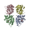
- PDB-3sr7: Crystal structure of S. mutans isopentenyl pyrophosphate isomerase -
+ Open data

- Basic information
| Entry | Database: PDB / ID: 3sr7 | ||||||
|---|---|---|---|---|---|---|---|
| Title | Crystal structure of S. mutans isopentenyl pyrophosphate isomerase | ||||||
 Components Components | Isopentenyl-diphosphate delta-isomerase | ||||||
 Keywords Keywords | ISOMERASE / isopentenyl pyrophosphate isomerase / Tim-barrel | ||||||
| Function / homology |  Function and homology information Function and homology informationisopentenyl-diphosphate Delta-isomerase / isopentenyl-diphosphate delta-isomerase activity / isoprenoid biosynthetic process / NADPH binding / FMN binding / oxidoreductase activity / magnesium ion binding / cytoplasm Similarity search - Function | ||||||
| Biological species |  Streptococcus mutans (bacteria) Streptococcus mutans (bacteria) | ||||||
| Method |  X-RAY DIFFRACTION / SYNCHROTRON / SAD / Resolution: 2.036 Å X-RAY DIFFRACTION / SYNCHROTRON / SAD / Resolution: 2.036 Å | ||||||
 Authors Authors | Liu, Y.H. / Fu, T.M. / Liu, X. / Su, X.D. | ||||||
 Citation Citation | Journal: To be Published Title: Crystal structure of S. mutans isopentenyl pyrophosphate isomerase Authors: Liu, Y.H. / Fu, T.M. / Liu, X. / Su, X.D. | ||||||
| History |
|
- Structure visualization
| Structure viewer | Molecule: MolmilJmol/JSmol |
|---|
- Downloads & links
-Download
| PDBx/mmCIF format | 3sr7.cif.gz | 220.4 KB | Display | PDBx/mmCIF format |
|---|---|---|---|---|
| PDB format | pdb3sr7.ent.gz | 174 KB | Display | PDB format |
| PDBx/mmJSON format | 3sr7.json.gz | Tree view | PDBx/mmJSON format | |
| Others |  Other downloads Other downloads |
-Validation report
| Arichive directory | https://data.pdbj.org/pub/pdb/validation_reports/sr/3sr7ftp://data.pdbj.org/pub/pdb/validation_reports/sr/3sr7 | HTTPS FTP |
|---|
-Related structure data
| Similar structure data |
|---|


-Links
 PDBj
PDBj
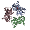
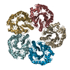
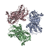
- Assembly
| Deposited unit | 
| ||||||||
|---|---|---|---|---|---|---|---|---|---|
| 1 |
| ||||||||
| Unit cell |
|
-Components
| #1: Protein | Mass: 40908.633 Da / Num. of mol.: 4 Source method: isolated from a genetically manipulated source Source: (gene. exp.) Streptococcus mutans (bacteria) / Strain: UA159 / Gene: fni, SMU_939 / Plasmid: pET28a / Production host: References: UniProt: Q8DUI9, isopentenyl-diphosphate Delta-isomerase #2: Chemical | ChemComp-PO4 / |   Mass: 94.971 Da / Num. of mol.: 1 / Source method: obtained synthetically / Formula: PO4 Mass: 94.971 Da / Num. of mol.: 1 / Source method: obtained synthetically / Formula: PO4#3: Water | ChemComp-HOH / |  Mass: 18.015 Da / Num. of mol.: 153 / Source method: isolated from a natural source / Formula: H2O Mass: 18.015 Da / Num. of mol.: 153 / Source method: isolated from a natural source / Formula: H2O |
|---|
-Experimental details
-Experiment
| Experiment | Method: X-RAY DIFFRACTION / Number of used crystals: 1 |
|---|
- Sample preparation
| Crystal | Density Matthews: 1.83 Å3/Da / Density % sol: 32.81 % |
|---|---|
| Crystal grow | Temperature: 293 K / Method: vapor diffusion, sitting drop / pH: 8 Details: 5mM MgCl2, 5mM MnCl2, 5mM CaCl2, 0.1M Tris HCl pH 8.0, 5% (w/v) PEG 8000, VAPOR DIFFUSION, SITTING DROP, temperature 293K |
-Data collection
| Diffraction | Mean temperature: 100 K |
|---|---|
| Diffraction source | Source: SYNCHROTRON / Site: Photon Factory  / Beamline: AR-NE3A / Wavelength: 0.9761 Å / Beamline: AR-NE3A / Wavelength: 0.9761 Å |
| Detector | Type: ADSC QUANTUM 270 / Detector: CCD / Date: Feb 6, 2010 |
| Radiation | Protocol: SINGLE WAVELENGTH / Monochromatic (M) / Laue (L): M / Scattering type: x-ray |
| Radiation wavelength | Wavelength: 0.9761 Å / Relative weight: 1 |
| Reflection | Resolution: 2.036→45.27 Å / Num. all: 75654 / Num. obs: 74368 / % possible obs: 98.3 % / Observed criterion σ(F): 2 / Observed criterion σ(I): 2 / Redundancy: 5 % / Rmerge(I) obs: 0.073 / Rsym value: 0.081 / Net I/σ(I): 10.6 |
| Reflection shell | Resolution: 2.04→2.15 Å / Redundancy: 4.9 % / Rmerge(I) obs: 0.56 / Mean I/σ(I) obs: 2.5 / Num. unique all: 10734 / Rsym value: 0.63 / % possible all: 97.5 |
- Processing
| Software |
| |||||||||||||||||||||||||||||||||||||||||||||||||||||||||||||||||||||||||||||||||||||||||||||||||||||||||
|---|---|---|---|---|---|---|---|---|---|---|---|---|---|---|---|---|---|---|---|---|---|---|---|---|---|---|---|---|---|---|---|---|---|---|---|---|---|---|---|---|---|---|---|---|---|---|---|---|---|---|---|---|---|---|---|---|---|---|---|---|---|---|---|---|---|---|---|---|---|---|---|---|---|---|---|---|---|---|---|---|---|---|---|---|---|---|---|---|---|---|---|---|---|---|---|---|---|---|---|---|---|---|---|---|---|---|
| Refinement | Method to determine structure: SAD / Resolution: 2.036→41.921 Å / SU ML: 0.35 / σ(F): 1.34 / Phase error: 34 / Stereochemistry target values: ML
| |||||||||||||||||||||||||||||||||||||||||||||||||||||||||||||||||||||||||||||||||||||||||||||||||||||||||
| Solvent computation | Shrinkage radii: 0.95 Å / VDW probe radii: 1.2 Å / Solvent model: FLAT BULK SOLVENT MODEL / Bsol: 63.184 Å2 / ksol: 0.379 e/Å3 | |||||||||||||||||||||||||||||||||||||||||||||||||||||||||||||||||||||||||||||||||||||||||||||||||||||||||
| Displacement parameters |
| |||||||||||||||||||||||||||||||||||||||||||||||||||||||||||||||||||||||||||||||||||||||||||||||||||||||||
| Refinement step | Cycle: LAST / Resolution: 2.036→41.921 Å
| |||||||||||||||||||||||||||||||||||||||||||||||||||||||||||||||||||||||||||||||||||||||||||||||||||||||||
| Refine LS restraints |
| |||||||||||||||||||||||||||||||||||||||||||||||||||||||||||||||||||||||||||||||||||||||||||||||||||||||||
| LS refinement shell |
|
