 Movie
Movie Controller

[English] 日本語
 Yorodumi
- PDB-3fok: Crystal Structure of Cgl0159 From Corynebacterium glutamicum (Bre... -
+ Open data

- Basic information
| Entry | Database: PDB / ID: 3fok | ||||||
|---|---|---|---|---|---|---|---|
| Title | Crystal Structure of Cgl0159 From Corynebacterium glutamicum (Brevibacterium flavum). Northeast Structural Genomics Target CgR115 | ||||||
 Components Components | uncharacterized protein Cgl0159 | ||||||
 Keywords Keywords | structural genomics / unknown function / Cgl0159 / Brevibacterium flavum. / PSI-2 / Protein Structure Initiative / Northeast Structural Genomics Consortium / NESG | ||||||
| Function / homology | : / Cgl0159-like / Aldolase class I / Aldolase-type TIM barrel / TIM Barrel / Alpha-Beta Barrel / Alpha Beta / Cgl0159-like domain-containing protein Function and homology information Function and homology information | ||||||
| Biological species |  Corynebacterium glutamicum (bacteria) Corynebacterium glutamicum (bacteria) | ||||||
| Method |  X-RAY DIFFRACTION / SYNCHROTRON / SAD / Resolution: 2.5 Å X-RAY DIFFRACTION / SYNCHROTRON / SAD / Resolution: 2.5 Å | ||||||
 Authors Authors | Seetharaman, J. / Neely, H. / Wang, H. / Janjua, H. / Foote, E.L. / Xiao, R. / Everett, J.K. / Acton, T.B. / Rost, B. / Montelione, G.T. ...Seetharaman, J. / Neely, H. / Wang, H. / Janjua, H. / Foote, E.L. / Xiao, R. / Everett, J.K. / Acton, T.B. / Rost, B. / Montelione, G.T. / Hunt, J.F. / Tong, L. / Northeast Structural Genomics Consortium (NESG) | ||||||
 Citation Citation | Journal: To be Published Title: Crystal Structure of Cgl0159 From Corynebacterium glutamicum (Brevibacterium flavum). Northeast Structural Genomics Target CgR115 Authors: Seetharaman, J. / Neely, H. / Wang, H. / Janjua, H. / Foote, E.L. / Xiao, R. / Everett, J.K. / Acton, T.B. / Rost, B. / Montelione, G.T. / Hunt, J.F. / Tong, L. | ||||||
| History |
|
- Structure visualization
| Structure viewer | Molecule: MolmilJmol/JSmol |
|---|
- Downloads & links
-Download
| PDBx/mmCIF format | 3fok.cif.gz | 553.7 KB | Display | PDBx/mmCIF format |
|---|---|---|---|---|
| PDB format | pdb3fok.ent.gz | 463.2 KB | Display | PDB format |
| PDBx/mmJSON format | 3fok.json.gz | Tree view | PDBx/mmJSON format | |
| Others |  Other downloads Other downloads |
-Validation report
| Arichive directory | https://data.pdbj.org/pub/pdb/validation_reports/fo/3fokftp://data.pdbj.org/pub/pdb/validation_reports/fo/3fok | HTTPS FTP |
|---|
-Related structure data
| Similar structure data | |
|---|---|
| Other databases |


-Links
 PDBj
PDBj- Assembly
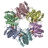
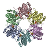
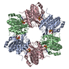
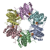
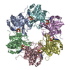
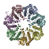
| Deposited unit | 
| ||||||||
|---|---|---|---|---|---|---|---|---|---|
| 1 | 
| ||||||||
| 2 | 
| ||||||||
| Unit cell |
| ||||||||
| Details | pentamer |
-Components
| #1: Protein | Mass: 33617.855 Da / Num. of mol.: 10 Source method: isolated from a genetically manipulated source Source: (gene. exp.) Corynebacterium glutamicum (bacteria) / Gene: Cgl0159, cg0198 / Production host: #2: Water | ChemComp-HOH / |  Mass: 18.015 Da / Num. of mol.: 715 / Source method: isolated from a natural source / Formula: H2O Mass: 18.015 Da / Num. of mol.: 715 / Source method: isolated from a natural source / Formula: H2OHas protein modification | Y | |
|---|
-Experimental details
-Experiment
| Experiment | Method: X-RAY DIFFRACTION / Number of used crystals: 1 |
|---|
- Sample preparation
| Crystal | Density Matthews: 2.51 Å3/Da / Density % sol: 51.04 % |
|---|---|
| Crystal grow | Temperature: 293 K / Method: vapor diffusion, sitting drop / pH: 4.5 Details: 0.2M NH4CL, 10% PEG 3350, , pH 4.5, VAPOR DIFFUSION, SITTING DROP, temperature 293K |
-Data collection
| Diffraction | Mean temperature: 100 K |
|---|---|
| Diffraction source | Source: SYNCHROTRON / Site: NSLS  / Beamline: X4A / Wavelength: 0.979 Å / Beamline: X4A / Wavelength: 0.979 Å |
| Detector | Type: ADSC QUANTUM 4 / Detector: CCD / Date: Nov 15, 2008 |
| Radiation | Protocol: SINGLE WAVELENGTH / Monochromatic (M) / Laue (L): M / Scattering type: x-ray |
| Radiation wavelength | Wavelength: 0.979 Å / Relative weight: 1 |
| Reflection | Resolution: 2.5→50 Å / Num. obs: 256002 / % possible obs: 99.1 % / Observed criterion σ(F): 0 / Observed criterion σ(I): 0 / Redundancy: 3.1 % / Biso Wilson estimate: 27.5 Å2 / Rmerge(I) obs: 0.106 / Net I/σ(I): 11.2 |
| Reflection shell | Resolution: 2.5→2.59 Å / Redundancy: 3 % / Rmerge(I) obs: 0.44 / Mean I/σ(I) obs: 10 / Num. unique all: 25191 / % possible all: 96.9 |
- Processing
| Software |
| ||||||||||||||||||||
|---|---|---|---|---|---|---|---|---|---|---|---|---|---|---|---|---|---|---|---|---|---|
| Refinement | Method to determine structure: SAD / Resolution: 2.5→39 Å / Rfactor Rfree error: 0.003 / Data cutoff high absF: 59317.31 / Data cutoff low absF: 0 / Isotropic thermal model: RESTRAINED / Cross valid method: THROUGHOUT / σ(F): 0 / Stereochemistry target values: Engh & Huber / Details: BULK SOLVENT MODEL USED
| ||||||||||||||||||||
| Solvent computation | Solvent model: FLAT MODEL / Bsol: 32.0521 Å2 / ksol: 0.4 e/Å3 | ||||||||||||||||||||
| Displacement parameters | Biso mean: 28.1 Å2
| ||||||||||||||||||||
| Refine analyze |
| ||||||||||||||||||||
| Refinement step | Cycle: LAST / Resolution: 2.5→39 Å
| ||||||||||||||||||||
| Refine LS restraints |
| ||||||||||||||||||||
| LS refinement shell | Resolution: 2.5→2.66 Å / Rfactor Rfree error: 0.008 / Total num. of bins used: 6
| ||||||||||||||||||||
| Xplor file |
|
