 Movie
Movie Controller

[English] 日本語
 Yorodumi
- PDB-3smd: Crystal structure of a mut/nudix family protein from bacillus thu... -
+ Open data

- Basic information
| Entry | Database: PDB / ID: 3smd | |||||||||
|---|---|---|---|---|---|---|---|---|---|---|
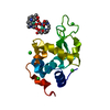
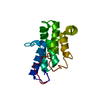
| Title | Crystal structure of a mut/nudix family protein from bacillus thuringiensis | |||||||||
 Components Components | MutT/NUDIX family protein | |||||||||
 Keywords Keywords | Structural Genomics / Unknown Function / PSI-2 / Protein Structure Initiative / New York SGX Research Center for Structural Genomics / NYSGXRC / Mut/NUDIX protein / Protein Structure Initiative II(PSI II) / 11181d / Hydrolase | |||||||||
| Function / homology |  Function and homology information Function and homology informationHydrolases; Acting on acid anhydrides; In phosphorus-containing anhydrides / hydrolase activity / membrane Similarity search - Function | |||||||||
| Biological species |  | |||||||||
| Method |  X-RAY DIFFRACTION / SYNCHROTRON / SAD / Resolution: 1.76 Å X-RAY DIFFRACTION / SYNCHROTRON / SAD / Resolution: 1.76 Å | |||||||||
 Authors Authors | Palani, K. / Kumaran, D. / Burley, S.K. / Swaminathan, S. / New York SGX Research Center for Structural Genomics (NYSGXRC) | |||||||||
 Citation Citation | Journal: To be Published Title: Crystal structure of a mut/nudix family protein from bacillus thuringiensis Authors: Palani, K. / Kumaran, D. / Burley, S.K. / Swaminathan, S. | |||||||||
| History |
|
- Structure visualization
| Structure viewer | Molecule: MolmilJmol/JSmol |
|---|
- Downloads & links
-Download
| PDBx/mmCIF format | 3smd.cif.gz | 42.5 KB | Display | PDBx/mmCIF format |
|---|---|---|---|---|
| PDB format | pdb3smd.ent.gz | 29 KB | Display | PDB format |
| PDBx/mmJSON format | 3smd.json.gz | Tree view | PDBx/mmJSON format | |
| Others |  Other downloads Other downloads |
-Validation report
| Summary document | 3smd_validation.pdf.gz | 423.6 KB | Display | wwPDB validaton report |
|---|---|---|---|---|
| Full document | 3smd_full_validation.pdf.gz | 424.1 KB | Display | |
| Data in XML | 3smd_validation.xml.gz | 8.5 KB | Display | |
| Data in CIF | 3smd_validation.cif.gz | 11.4 KB | Display | |
| Arichive directory | https://data.pdbj.org/pub/pdb/validation_reports/sm/3smdftp://data.pdbj.org/pub/pdb/validation_reports/sm/3smd | HTTPS FTP |
-Related structure data
| Similar structure data | |
|---|---|
| Other databases |

-Links
 PDBj
PDBj

- Assembly
| Deposited unit | 
| ||||||||
|---|---|---|---|---|---|---|---|---|---|
| 1 |
| ||||||||
| 2 | 
| ||||||||
| Unit cell |
|
-Components
| #1: Protein | Mass: 17589.748 Da / Num. of mol.: 1 Source method: isolated from a genetically manipulated source Source: (gene. exp.) |
|---|---|
| #2: Water | ChemComp-HOH /  Mass: 18.015 Da / Num. of mol.: 124 / Source method: isolated from a natural source / Formula: H2O Mass: 18.015 Da / Num. of mol.: 124 / Source method: isolated from a natural source / Formula: H2O |
| Has protein modification | Y |
-Experimental details
-Experiment
| Experiment | Method: X-RAY DIFFRACTION / Number of used crystals: 1 |
|---|
- Sample preparation
| Crystal | Density Matthews: 1.98 Å3/Da / Density % sol: 37.98 % |
|---|---|
| Crystal grow | Temperature: 298 K / Method: vapor diffusion, sitting drop / pH: 7.5 Details: 0.1M HEPES-Na, 10% isopropanal, 20% PEG4000 , pH 7.5, VAPOR DIFFUSION, SITTING DROP, temperature 298.0K |
-Data collection
| Diffraction | Mean temperature: 100 K |
|---|---|
| Diffraction source | Source: SYNCHROTRON / Site: NSLS  / Beamline: X12C / Wavelength: 0.9205 Å / Beamline: X12C / Wavelength: 0.9205 Å |
| Detector | Type: ADSC QUANTUM 210 / Detector: CCD / Date: Aug 17, 2008 / Details: mirrors |
| Radiation | Monochromator: Si(III) / Protocol: SINGLE WAVELENGTH / Monochromatic (M) / Laue (L): M / Scattering type: x-ray |
| Radiation wavelength | Wavelength: 0.9205 Å / Relative weight: 1 |
| Reflection | Resolution: 1.76→50 Å / Num. all: 14215 / Num. obs: 14215 / % possible obs: 99.5 % / Observed criterion σ(F): 0 / Observed criterion σ(I): 0 / Redundancy: 21.1 % / Biso Wilson estimate: 15.2 Å2 / Rmerge(I) obs: 0.042 / Net I/σ(I): 28 |
| Reflection shell | Resolution: 1.76→1.82 Å / Redundancy: 18.9 % / Rmerge(I) obs: 0.447 / Mean I/σ(I) obs: 4 / Num. unique all: 1324 / % possible all: 94.9 |
- Processing
| Software |
| |||||||||||||||||||||||||
|---|---|---|---|---|---|---|---|---|---|---|---|---|---|---|---|---|---|---|---|---|---|---|---|---|---|---|
| Refinement | Method to determine structure: SAD / Resolution: 1.76→40.4 Å / Rfactor Rfree error: 0.01 / Data cutoff high absF: 123240.09 / Data cutoff low absF: 0 / Isotropic thermal model: RESTRAINED / Cross valid method: THROUGHOUT / σ(F): 0 / Stereochemistry target values: Engh & Huber
| |||||||||||||||||||||||||
| Solvent computation | Solvent model: FLAT MODEL / Bsol: 46.869 Å2 / ksol: 0.357741 e/Å3 | |||||||||||||||||||||||||
| Displacement parameters | Biso mean: 24.1 Å2
| |||||||||||||||||||||||||
| Refine analyze |
| |||||||||||||||||||||||||
| Refinement step | Cycle: LAST / Resolution: 1.76→40.4 Å
| |||||||||||||||||||||||||
| Refine LS restraints |
| |||||||||||||||||||||||||
| Refine LS restraints NCS | NCS model details: NONE | |||||||||||||||||||||||||
| LS refinement shell | Resolution: 1.76→1.87 Å / Rfactor Rfree error: 0.029 / Total num. of bins used: 6
| |||||||||||||||||||||||||
| Xplor file |
|
