 Movie
Movie Controller


+ Open data

- Basic information
| Entry | Database: PDB / ID: 3sil | ||||||
|---|---|---|---|---|---|---|---|
| Title | SIALIDASE FROM SALMONELLA TYPHIMURIUM | ||||||
 Components Components | SIALIDASE | ||||||
 Keywords Keywords | GLYCOSIDASE / HYDROLASE | ||||||
| Function / homology |  Function and homology information Function and homology informationganglioside catabolic process / oligosaccharide catabolic process / exo-alpha-sialidase / exo-alpha-sialidase activity / intracellular membrane-bounded organelle / membrane / cytoplasm Similarity search - Function | ||||||
| Biological species |  Salmonella typhimurium (bacteria) Salmonella typhimurium (bacteria) | ||||||
| Method |  X-RAY DIFFRACTION / SYNCHROTRON / Resolution: 1.05 Å X-RAY DIFFRACTION / SYNCHROTRON / Resolution: 1.05 Å | ||||||
 Authors Authors | Garman, E.F. / Sheldrick, G.M. | ||||||
 Citation Citation | Journal: To be Published Title: An Enzyme at Atomic Resolution: The 1.05 A Structure of Salmonella Typhimurium Neuraminidase (Sialidase) Authors: Garman, E.F. / Wouters, J. / Schneider, T.R. / Vimr, E.R. / Laver, W.G. / Sheldrick, G.M. #1: Journal: J.Mol.Biol. / Year: 1996Title: The Structures of Salmonella Typhimurium Lt2 Neuraminidase and its Complexes with Three Inhibitors at High Resolution Authors: Crennell, S.J. / Garman, E.F. / Philippon, C. / Vasella, A. / Laver, W.G. / Vimr, E.R. / Taylor, G.L. #2: Journal: Proc.Natl.Acad.Sci.USA / Year: 1993Title: Crystal Structure of a Bacterial Sialidase (from Salmonella Typhimurium Lt2) Shows the Same Fold as an Influenza Virus Neuraminidase Authors: Crennell, S.J. / Garman, E.F. / Laver, W.G. / Vimr, E.R. / Taylor, G.L. | ||||||
| History |
|
- Structure visualization
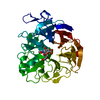
| Structure viewer | Molecule: MolmilJmol/JSmol |
|---|
- Downloads & links
-Download
| PDBx/mmCIF format | 3sil.cif.gz | 190.7 KB | Display | PDBx/mmCIF format |
|---|---|---|---|---|
| PDB format | pdb3sil.ent.gz | 150.6 KB | Display | PDB format |
| PDBx/mmJSON format | 3sil.json.gz | Tree view | PDBx/mmJSON format | |
| Others |  Other downloads Other downloads |
-Validation report
| Summary document | 3sil_validation.pdf.gz | 392.2 KB | Display | wwPDB validaton report |
|---|---|---|---|---|
| Full document | 3sil_full_validation.pdf.gz | 396 KB | Display | |
| Data in XML | 3sil_validation.xml.gz | 9.9 KB | Display | |
| Data in CIF | 3sil_validation.cif.gz | 18.1 KB | Display | |
| Arichive directory | https://data.pdbj.org/pub/pdb/validation_reports/si/3silftp://data.pdbj.org/pub/pdb/validation_reports/si/3sil | HTTPS FTP |
-Related structure data
| Related structure data |  2silS S: Starting model for refinement |
|---|---|
| Similar structure data |


-Links
 PDBj
PDBj

- Assembly
| Deposited unit | 
| ||||||||
|---|---|---|---|---|---|---|---|---|---|
| 1 |
| ||||||||
| Unit cell |
|
-Components
| #1: Protein | Mass: 41791.555 Da / Num. of mol.: 1 / Mutation: RESIDUE MET 1 WAS EXCISED BY ESCHERICHIA COLI Source method: isolated from a genetically manipulated source Source: (gene. exp.) Salmonella typhimurium (bacteria) / Strain: LT2 / Gene: NANH / Variant: TA263, PROTOTROPH / Plasmid: PSX62 / Gene (production host): NANH / Production host: | ||||
|---|---|---|---|---|---|
| #2: Chemical | ChemComp-K /   Mass: 39.098 Da / Num. of mol.: 1 / Source method: obtained synthetically / Formula: K Mass: 39.098 Da / Num. of mol.: 1 / Source method: obtained synthetically / Formula: K | ||||
| #3: Chemical | ChemComp-PO4 /   Mass: 94.971 Da / Num. of mol.: 1 / Source method: obtained synthetically / Formula: PO4 Mass: 94.971 Da / Num. of mol.: 1 / Source method: obtained synthetically / Formula: PO4 | ||||
| #4: Chemical | ChemComp-GOL /   Mass: 92.094 Da / Num. of mol.: 6 / Source method: obtained synthetically / Formula: C3H8O3 Mass: 92.094 Da / Num. of mol.: 6 / Source method: obtained synthetically / Formula: C3H8O3#5: Water | ChemComp-HOH / |  Mass: 18.015 Da / Num. of mol.: 561 / Source method: isolated from a natural source / Formula: H2O Mass: 18.015 Da / Num. of mol.: 561 / Source method: isolated from a natural source / Formula: H2OHas protein modification | Y | |
-Experimental details
-Experiment
| Experiment | Method: X-RAY DIFFRACTION / Number of used crystals: 1 |
|---|
- Sample preparation
| Crystal | Density Matthews: 2.1 Å3/Da / Density % sol: 41.4 % |
|---|---|
| Crystal grow | pH: 7.86 / Details: pH 7.86 |
-Data collection
| Diffraction | Mean temperature: 100 K |
|---|---|
| Diffraction source | Source: SYNCHROTRON / Site: EMBL/DESY, HAMBURG  / Beamline: BW7B / Wavelength: 0.862 / Beamline: BW7B / Wavelength: 0.862 |
| Detector | Type: MARRESEARCH / Detector: IMAGE PLATE / Date: May 1, 1995 |
| Radiation | Monochromatic (M) / Laue (L): M / Scattering type: x-ray |
| Radiation wavelength | Wavelength: 0.862 Å / Relative weight: 1 |
| Reflection | Resolution: 1.05→11 Å / Num. obs: 148478 / % possible obs: 90.1 % / Redundancy: 3.6 % / Rmerge(I) obs: 0.056 / Net I/σ(I): 8.5 |
| Reflection shell | Resolution: 1.05→1.15 Å / Redundancy: 2.6 % / Rmerge(I) obs: 0.346 / Mean I/σ(I) obs: 3.4 / % possible all: 89.8 |
- Processing
| Software |
| |||||||||||||||||||||||||||||||||
|---|---|---|---|---|---|---|---|---|---|---|---|---|---|---|---|---|---|---|---|---|---|---|---|---|---|---|---|---|---|---|---|---|---|---|
| Refinement | Starting model: PDB ENTRY 2SIL Highest resolution: 1.05 Å / Num. parameters: 33361 / Num. restraintsaints: 41703 / Cross valid method: FREE R / σ(F): 0 / Stereochemistry target values: ENGH AND HUBER / Details: ANISOTROPIC REFINEMENT REDUCED FREE R (NO CUTOFF).
| |||||||||||||||||||||||||||||||||
| Solvent computation | Solvent model: MOEWS & KRETSINGER, J.MOL.BIOL. (1973) 91: 201-228 | |||||||||||||||||||||||||||||||||
| Refine analyze | Num. disordered residues: 47 / Occupancy sum hydrogen: 2797.8 / Occupancy sum non hydrogen: 3526.4 | |||||||||||||||||||||||||||||||||
| Refinement step | Cycle: LAST / Highest resolution: 1.05 Å
| |||||||||||||||||||||||||||||||||
| Refine LS restraints |
|
