- PDB-3sgi: Crystal structure of DNA ligase A BRCT domain deleted mutant of M... -
+
Open data
ID or keywords:
Loading...
-
Basic information
Entry
Database: PDB / ID: 3sgi
Title
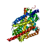
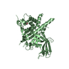
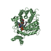
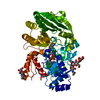
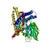
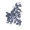
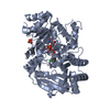
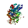
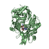
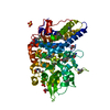
Crystal structure of DNA ligase A BRCT domain deleted mutant of Mycobacterium tuberculosis
Components
DNA ligase
Keywords
LIGASE / NAD depndent DNA ligase A
Function / homology
Function and homology information
DNA ligase (NAD+) / DNA ligase (NAD+) activity / peptidoglycan-based cell wall / DNA replication / DNA repair / magnesium ion binding / metal ion binding / plasma membrane / cytosol Similarity search - Function
Zinc-finger, NAD-dependent DNA ligase C4-type / NAD-dependent DNA ligase C4 zinc finger domain / NAD-dependent DNA ligase, active site / NAD-dependent DNA ligase, conserved site / NAD-dependent DNA ligase signature 1. / NAD-dependent DNA ligase signature 2. / NAD-dependent DNA ligase / NAD-dependent DNA ligase, OB-fold / NAD-dependent DNA ligase, adenylation / NAD-dependent DNA ligase, N-terminal ...Zinc-finger, NAD-dependent DNA ligase C4-type / NAD-dependent DNA ligase C4 zinc finger domain / NAD-dependent DNA ligase, active site / NAD-dependent DNA ligase, conserved site / NAD-dependent DNA ligase signature 1. / NAD-dependent DNA ligase signature 2. / NAD-dependent DNA ligase / NAD-dependent DNA ligase, OB-fold / NAD-dependent DNA ligase, adenylation / NAD-dependent DNA ligase, N-terminal / NAD-dependent DNA ligase adenylation domain / NAD-dependent DNA ligase OB-fold domain / Ligase N family / DisA/LigA, helix-hairpin-helix motif / Helix-hairpin-helix motif / DNA ligase/mRNA capping enzyme / Helix hairpin bin / RuvA domain 2-like / BRCA1 C Terminus (BRCT) domain / breast cancer carboxy-terminal domain / D-amino Acid Aminotransferase; Chain A, domain 1 / BRCT domain profile. / BRCT domain / BRCT domain superfamily / Nucleic acid-binding proteins / OB fold (Dihydrolipoamide Acetyltransferase, E2P) / Helix Hairpins / Nucleic acid-binding, OB-fold / Beta Barrel / 2-Layer Sandwich / Orthogonal Bundle / Mainly Beta / Mainly Alpha / Alpha Beta Similarity search - Domain/homology
Journal: To be Published Title: Crystal Structure of Domain deleted mutant of Mycobacterium tuberculosis NAD+ dependent DNA ligase capture the AMP cofactor in a new state Authors: Kukshal, V. / Ravishankar, R.
In the structure databanks used in Yorodumi, some data are registered as the other names, "COVID-19 virus" and "2019-nCoV". Here are the details of the virus and the list of structure data.
Jan 31, 2019. EMDB accession codes are about to change! (news from PDBe EMDB page)
EMDB accession codes are about to change! (news from PDBe EMDB page)
The allocation of 4 digits for EMDB accession codes will soon come to an end. Whilst these codes will remain in use, new EMDB accession codes will include an additional digit and will expand incrementally as the available range of codes is exhausted. The current 4-digit format prefixed with “EMD-” (i.e. EMD-XXXX) will advance to a 5-digit format (i.e. EMD-XXXXX), and so on. It is currently estimated that the 4-digit codes will be depleted around Spring 2019, at which point the 5-digit format will come into force.
The EM Navigator/Yorodumi systems omit the EMD- prefix.
Related info.:Q: What is EMD? / ID/Accession-code notation in Yorodumi/EM Navigator
Yorodumi is a browser for structure data from EMDB, PDB, SASBDB, etc.
This page is also the successor to EM Navigator detail page, and also detail information page/front-end page for Omokage search.
The word "yorodu" (or yorozu) is an old Japanese word meaning "ten thousand". "mi" (miru) is to see.
Related info.:EMDB / PDB / SASBDB / Comparison of 3 databanks / Yorodumi Search / Aug 31, 2016. New EM Navigator & Yorodumi / Yorodumi Papers / Jmol/JSmol / Function and homology information / Changes in new EM Navigator and Yorodumi
 Movie
Movie Controller

 Yorodumi
 Open data
 Basic information
 Components
 Keywords
 Function and homology information
 Mycobacterium tuberculosis (bacteria)
 X-RAY DIFFRACTION /
  Authors
 Citation
 Structure visualization
 Downloads & links
 Other downloads
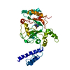
 PDBj
PDBj


 Assembly
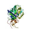

 Mass: 347.221 Da / Num. of mol.: 1 / Source method: obtained synthetically / Formula: C10H14N5O7P / Comment: AMP*YM
 Mass: 18.015 Da / Num. of mol.: 23 / Source method: isolated from a natural source / Formula: H2O
 Sample preparation
 Processing