 Movie
Movie Controller

[English] 日本語
 Yorodumi
- PDB-3sgg: Crystal structure of a putative hydrolase (BT_2193) from Bacteroi... -
+ Open data

- Basic information
| Entry | Database: PDB / ID: 3sgg | ||||||
|---|---|---|---|---|---|---|---|
| Title | Crystal structure of a putative hydrolase (BT_2193) from Bacteroides thetaiotaomicron VPI-5482 at 1.25 A resolution | ||||||
 Components Components | Hypothetical hydrolase | ||||||
 Keywords Keywords | HYDROLASE / 7-stranded beta/alpha barrel / Structural Genomics / Joint Center for Structural Genomics / JCSG / Protein Structure Initiative / PSI-BIOLOGY | ||||||
| Function / homology |  Function and homology information Function and homology informationGxGYxYP glycoside hydrolase, C-terminal domain / GxGYxYP putative glycoside hydrolase, C-terminal / GxGYxYP putative glycoside hydrolase, N-terminal / GxGYxYP putative glycoside hydrolase, C-terminal domain superfamily / : / : / GxGYxYP putative glycoside hydrolase C-terminal domain / GxGYxYP_N 1st domain / GxGYxYP_N second domain / GxGYxYP third domain ...GxGYxYP glycoside hydrolase, C-terminal domain / GxGYxYP putative glycoside hydrolase, C-terminal / GxGYxYP putative glycoside hydrolase, N-terminal / GxGYxYP putative glycoside hydrolase, C-terminal domain superfamily / : / : / GxGYxYP putative glycoside hydrolase C-terminal domain / GxGYxYP_N 1st domain / GxGYxYP_N second domain / GxGYxYP third domain / Prokaryotic membrane lipoprotein lipid attachment site profile. / TIM Barrel / Alpha-Beta Barrel / Alpha Beta Similarity search - Domain/homology | ||||||
| Biological species |  Bacteroides thetaiotaomicron (bacteria) Bacteroides thetaiotaomicron (bacteria) | ||||||
| Method |  X-RAY DIFFRACTION / SYNCHROTRON / MAD / Resolution: 1.25 Å X-RAY DIFFRACTION / SYNCHROTRON / MAD / Resolution: 1.25 Å | ||||||
 Authors Authors | Joint Center for Structural Genomics (JCSG) | ||||||
 Citation Citation | Journal: To be published Title: Crystal structure of a Hypothetical hydrolase (BT_2193) from Bacteroides thetaiotaomicron VPI-5482 at 1.25 A resolution Authors: Joint Center for Structural Genomics (JCSG) | ||||||
| History |
|
- Structure visualization
| Structure viewer | Molecule: MolmilJmol/JSmol |
|---|
- Downloads & links
-Download
| PDBx/mmCIF format | 3sgg.cif.gz | 252 KB | Display | PDBx/mmCIF format |
|---|---|---|---|---|
| PDB format | pdb3sgg.ent.gz | 200.6 KB | Display | PDB format |
| PDBx/mmJSON format | 3sgg.json.gz | Tree view | PDBx/mmJSON format | |
| Others |  Other downloads Other downloads |
-Validation report
| Arichive directory | https://data.pdbj.org/pub/pdb/validation_reports/sg/3sggftp://data.pdbj.org/pub/pdb/validation_reports/sg/3sgg | HTTPS FTP |
|---|
-Related structure data
| Similar structure data | |
|---|---|
| Other databases |

-Links
 PDBj
PDBj


- Assembly
| Deposited unit | 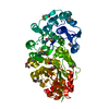
| ||||||||
|---|---|---|---|---|---|---|---|---|---|
| 1 |
| ||||||||
| Unit cell |
|
-Components
| #1: Protein | Mass: 60092.273 Da / Num. of mol.: 1 / Fragment: sequence database residues 23-557 Source method: isolated from a genetically manipulated source Source: (gene. exp.) Bacteroides thetaiotaomicron (bacteria)Gene: BT_2193 / Plasmid: SpeedET / Production host: | ||||||
|---|---|---|---|---|---|---|---|
| #2: Chemical | ChemComp-GOL /   Mass: 92.094 Da / Num. of mol.: 5 / Source method: obtained synthetically / Formula: C3H8O3 Mass: 92.094 Da / Num. of mol.: 5 / Source method: obtained synthetically / Formula: C3H8O3#3: Water | ChemComp-HOH / |  Mass: 18.015 Da / Num. of mol.: 550 / Source method: isolated from a natural source / Formula: H2O Mass: 18.015 Da / Num. of mol.: 550 / Source method: isolated from a natural source / Formula: H2OHas protein modification | Y | Sequence details | THE CONSTRUCT (RESIDUES 23-557) WAS EXPRESSED WITH A PURIFICATION TAG MGSDKIHHHHHHENLYFQG. THE TAG ...THE CONSTRUCT (RESIDUES 23-557) WAS EXPRESSED WITH A PURIFICATI | |
-Experimental details
-Experiment
| Experiment | Method: X-RAY DIFFRACTION / Number of used crystals: 1 |
|---|
- Sample preparation
| Crystal | Density Matthews: 2.16 Å3/Da / Density % sol: 43.06 % Description: DATA WERE SCALED USING XSCALE WITH FRIEDEL PAIRS KEPT AS SEPARATE WHEN COMPUTING R MERGE, COMPLETENESS AND Crystal grow | Temperature: 293 K / Method: vapor diffusion, sitting drop / pH: 5 | Details: 20.00% polyethylene glycol 6000, 0.1M sodium citrate pH 5.0, NANODROP, VAPOR DIFFUSION, SITTING DROP, temperature 293K |
|---|
-Data collection
| Diffraction | Mean temperature: 100 K | |||||||||||||||||||||||||||||||||||||||||||||||||||||||||||||||||||||||||||||
|---|---|---|---|---|---|---|---|---|---|---|---|---|---|---|---|---|---|---|---|---|---|---|---|---|---|---|---|---|---|---|---|---|---|---|---|---|---|---|---|---|---|---|---|---|---|---|---|---|---|---|---|---|---|---|---|---|---|---|---|---|---|---|---|---|---|---|---|---|---|---|---|---|---|---|---|---|---|---|
| Diffraction source | Source: SYNCHROTRON / Site: SSRL  / Beamline: BL11-1 / Wavelength: 0.91837,0.97941,0.97904 / Beamline: BL11-1 / Wavelength: 0.91837,0.97941,0.97904 | |||||||||||||||||||||||||||||||||||||||||||||||||||||||||||||||||||||||||||||
| Detector | Type: MARMOSAIC 325 mm CCD / Detector: CCD / Date: May 25, 2011 Details: Flat mirror (vertical focusing); single crystal Si(111) bent monochromator (ho rizontal focusing) | |||||||||||||||||||||||||||||||||||||||||||||||||||||||||||||||||||||||||||||
| Radiation | Monochromator: single crystal Si(111) bent / Protocol: MAD / Monochromatic (M) / Laue (L): M / Scattering type: x-ray | |||||||||||||||||||||||||||||||||||||||||||||||||||||||||||||||||||||||||||||
| Radiation wavelength |
| |||||||||||||||||||||||||||||||||||||||||||||||||||||||||||||||||||||||||||||
| Reflection | Resolution: 1.25→28.498 Å / Num. obs: 140636 / % possible obs: 97.3 % / Observed criterion σ(I): -3 / Biso Wilson estimate: 11.346 Å2 / Rmerge(I) obs: 0.041 / Net I/σ(I): 10.5 | |||||||||||||||||||||||||||||||||||||||||||||||||||||||||||||||||||||||||||||
| Reflection shell |
|
-Phasing
| Phasing | Method: MAD |
|---|
- Processing
| Software |
| ||||||||||||||||||||||||||||||||||||||||||||||||||||||||||||||||||||||||||||||||||||||||||||||||||||
|---|---|---|---|---|---|---|---|---|---|---|---|---|---|---|---|---|---|---|---|---|---|---|---|---|---|---|---|---|---|---|---|---|---|---|---|---|---|---|---|---|---|---|---|---|---|---|---|---|---|---|---|---|---|---|---|---|---|---|---|---|---|---|---|---|---|---|---|---|---|---|---|---|---|---|---|---|---|---|---|---|---|---|---|---|---|---|---|---|---|---|---|---|---|---|---|---|---|---|---|---|---|
| Refinement | Method to determine structure: MAD / Resolution: 1.25→28.498 Å / Cor.coef. Fo:Fc: 0.966 / Cor.coef. Fo:Fc free: 0.962 / Occupancy max: 1 / Occupancy min: 0.2 / SU B: 1.424 / SU ML: 0.028 / Cross valid method: THROUGHOUT / σ(F): 0 / ESU R: 0.046 / ESU R Free: 0.043 Stereochemistry target values: MAXIMUM LIKELIHOOD WITH PHASES Details: 1. HYDROGENS HAVE BEEN ADDED IN THE RIDING POSITIONS. 2. A MET-INHIBITION PROTOCOL WAS USED FOR SELENOMETHIONINE INCORPORATION DURING PROTEIN EXPRESSION. THE OCCUPANCY OF THE SE ATOMS IN THE ...Details: 1. HYDROGENS HAVE BEEN ADDED IN THE RIDING POSITIONS. 2. A MET-INHIBITION PROTOCOL WAS USED FOR SELENOMETHIONINE INCORPORATION DURING PROTEIN EXPRESSION. THE OCCUPANCY OF THE SE ATOMS IN THE MSE RESIDUES WAS REDUCED TO 0.75 FOR THE REDUCED SCATTERING POWER DUE TO PARTIAL S-MET INCORPORATION. 3. GLYCEROL (GOL) MODELED ARE PRESENT IN CRYO CONDITION. 4. ANISOTROPIC B-VALUE REFINEMENT WAS USED.
| ||||||||||||||||||||||||||||||||||||||||||||||||||||||||||||||||||||||||||||||||||||||||||||||||||||
| Solvent computation | Ion probe radii: 0.8 Å / Shrinkage radii: 0.8 Å / VDW probe radii: 1.2 Å / Solvent model: MASK | ||||||||||||||||||||||||||||||||||||||||||||||||||||||||||||||||||||||||||||||||||||||||||||||||||||
| Displacement parameters | Biso max: 51.94 Å2 / Biso mean: 15.7021 Å2 / Biso min: 5.2 Å2
| ||||||||||||||||||||||||||||||||||||||||||||||||||||||||||||||||||||||||||||||||||||||||||||||||||||
| Refinement step | Cycle: LAST / Resolution: 1.25→28.498 Å
| ||||||||||||||||||||||||||||||||||||||||||||||||||||||||||||||||||||||||||||||||||||||||||||||||||||
| Refine LS restraints |
| ||||||||||||||||||||||||||||||||||||||||||||||||||||||||||||||||||||||||||||||||||||||||||||||||||||
| LS refinement shell | Resolution: 1.25→1.282 Å / Total num. of bins used: 20
|
