Type: MARMOSAIC 325 mm CCD / Detector: CCD / Date: Jul 7, 2009 Details: Flat mirror (vertical focusing); single crystal Si(111) bent monochromator (ho rizontal focusing)
Radiation
Monochromator: single crystal Si(111) bent / Protocol: MAD / Monochromatic (M) / Laue (L): M / Scattering type: x-ray
Radiation wavelength
ID
Wavelength (Å)
Relative weight
1
0.97954
1
2
0.91837
1
3
0.97874
1
Reflection
Resolution: 1.85→36.25 Å / Num. obs: 16569 / % possible obs: 97.1 % / Observed criterion σ(I): -3 / Redundancy: 3.98 % / Biso Wilson estimate: 20.25 Å2 / Rmerge(I) obs: 0.097 / Net I/σ(I): 11.41
Reflection shell
Diffraction-ID: 1
Resolution (Å)
Redundancy (%)
Rmerge(I) obs
Mean I/σ(I) obs
Num. measured obs
Num. unique obs
% possible all
1.85-1.92
3.99
0.683
2.1
6715
1684
96.1
1.92-1.99
0.521
2.8
5932
1469
96.3
1.99-2.08
0.374
3.8
6565
1629
96.7
2.08-2.19
0.3
4.7
6587
1623
96.8
2.19-2.33
0.239
5.9
6740
1664
96.8
2.33-2.51
0.176
7.7
6698
1658
97.6
2.51-2.76
0.129
10.2
6652
1657
97.5
2.76-3.16
0.079
15.3
6712
1678
98.2
3.16-3.98
0.042
25.8
6647
1691
97.6
3.98-36.25
0.033
32.3
6742
1816
97.3
-
Phasing
Phasing
Method: MAD
-
Processing
Software
Name
Version
Classification
NB
MolProbity
3beta29
modelbuilding
PDB_EXTRACT
3.1
dataextraction
SHELX
phasing
SHARP
phasing
XSCALE
January30
datascaling
REFMAC
5.5.0110
refinement
XDS
datareduction
SHELXD
phasing
autoSHARP
phasing
Refinement
Method to determine structure: MAD / Resolution: 1.85→36.25 Å / Cor.coef. Fo:Fc: 0.962 / Cor.coef. Fo:Fc free: 0.945 / Occupancy max: 1 / Occupancy min: 0.5 / SU B: 5.588 / SU ML: 0.1 / Cross valid method: THROUGHOUT / σ(F): 0 / ESU R: 0.162 / ESU R Free: 0.136 Stereochemistry target values: MAXIMUM LIKELIHOOD WITH PHASES Details: 1. HYDROGENS HAVE BEEN ADDED IN THE RIDING POSITIONS. 2. ATOM RECORD CONTAINS SUM OF TLS AND RESIDUAL B FACTORS. 3. ANISOU RECORD CONTAINS SUM OF TLS AND RESIDUAL U FACTORS. 4. A MET- ...Details: 1. HYDROGENS HAVE BEEN ADDED IN THE RIDING POSITIONS. 2. ATOM RECORD CONTAINS SUM OF TLS AND RESIDUAL B FACTORS. 3. ANISOU RECORD CONTAINS SUM OF TLS AND RESIDUAL U FACTORS. 4. A MET-INHIBITION PROTOCOL WAS USED FOR SELENOMETHIONINE INCORPORATION DURING PROTEIN EXPRESSION. THE OCCUPANCY OF THE SE ATOMS IN THE MSE RESIDUES WAS REDUCED TO 0.75 FOR THE REDUCED SCATTERING POWER DUE TO PARTIAL S-MET INCORPORATION. 5. ANOMALOUS DIFFERENCE FOURIERS SUPPORT THE MODELING OF IODIDE (IOD) IONS. 6. 1,2-ETHANEDIOL (EDO) MOLECULES FROM THE CRYOPROTECTION SOLUTION ARE MODELED.
Rfactor
Num. reflection
% reflection
Selection details
Rfree
0.2018
854
5.2 %
RANDOM
Rwork
0.1693
-
-
-
obs
0.1711
16554
97.26 %
-
Solvent computation
Ion probe radii: 0.8 Å / Shrinkage radii: 0.8 Å / VDW probe radii: 1.4 Å / Solvent model: BABINET MODEL WITH MASK
In the structure databanks used in Yorodumi, some data are registered as the other names, "COVID-19 virus" and "2019-nCoV". Here are the details of the virus and the list of structure data.
Jan 31, 2019. EMDB accession codes are about to change! (news from PDBe EMDB page)
EMDB accession codes are about to change! (news from PDBe EMDB page)
The allocation of 4 digits for EMDB accession codes will soon come to an end. Whilst these codes will remain in use, new EMDB accession codes will include an additional digit and will expand incrementally as the available range of codes is exhausted. The current 4-digit format prefixed with “EMD-” (i.e. EMD-XXXX) will advance to a 5-digit format (i.e. EMD-XXXXX), and so on. It is currently estimated that the 4-digit codes will be depleted around Spring 2019, at which point the 5-digit format will come into force.
The EM Navigator/Yorodumi systems omit the EMD- prefix.
Related info.:Q: What is EMD? / ID/Accession-code notation in Yorodumi/EM Navigator
Yorodumi is a browser for structure data from EMDB, PDB, SASBDB, etc.
This page is also the successor to EM Navigator detail page, and also detail information page/front-end page for Omokage search.
The word "yorodu" (or yorozu) is an old Japanese word meaning "ten thousand". "mi" (miru) is to see.
Related info.:EMDB / PDB / SASBDB / Comparison of 3 databanks / Yorodumi Search / Aug 31, 2016. New EM Navigator & Yorodumi / Yorodumi Papers / Jmol/JSmol / Function and homology information / Changes in new EM Navigator and Yorodumi
 Movie
Movie Controller

 Yorodumi
 Open data
 Basic information
 Components
 Keywords
 Function and homology information
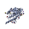
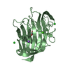
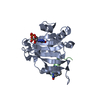
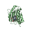
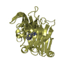
 Parabacteroides distasonis (bacteria)
 X-RAY DIFFRACTION /
  Authors
 Citation
 Structure visualization
 Downloads & links
 Other downloads
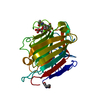


 PDBj
PDBj


 Assembly

 Mass: 126.904 Da / Num. of mol.: 3 / Source method: obtained synthetically / Formula: I
 Mass: 62.068 Da / Num. of mol.: 6 / Source method: obtained synthetically / Formula: C2H6O2
 Mass: 18.015 Da / Num. of mol.: 187 / Source method: isolated from a natural source / Formula: H2O
 Sample preparation
 / Beamline: BL11-1 / Wavelength: 0.97954,0.91837,0.97874
 Processing