 Movie
Movie Controller

[English] 日本語
 Yorodumi
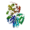
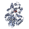
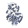
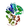
- PDB-3s44: Crystal Structure of Pasteurella multocida sialyltransferase M144... -
+ Open data

- Basic information
| Entry | Database: PDB / ID: 3s44 | ||||||
|---|---|---|---|---|---|---|---|
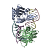
| Title | Crystal Structure of Pasteurella multocida sialyltransferase M144D mutant with CMP bound | ||||||
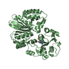
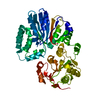
 Components Components | Alpha-2,3/2,6-sialyltransferase/sialidase | ||||||
 Keywords Keywords | TRANSFERASE / GT-B fold / Sialyltransferase | ||||||
| Function / homology |  Function and homology information Function and homology information | ||||||
| Biological species |  Pasteurella multocida (bacteria) Pasteurella multocida (bacteria) | ||||||
| Method |  X-RAY DIFFRACTION / SYNCHROTRON / MOLECULAR REPLACEMENT / Resolution: 1.45 Å X-RAY DIFFRACTION / SYNCHROTRON / MOLECULAR REPLACEMENT / Resolution: 1.45 Å | ||||||
 Authors Authors | Sugiarto, G. / Lau, K. / Li, Y. / Lim, S. / Ames, J.B. / Le, D.-T. / Fisher, A.J. / Chen, X. | ||||||
 Citation Citation | Journal: Acs Chem.Biol. / Year: 2012 Title: A Sialyltransferase Mutant with Decreased Donor Hydrolysis and Reduced Sialidase Activities for Directly Sialylating Lewis(x). Authors: Sugiarto, G. / Lau, K. / Qu, J. / Li, Y. / Lim, S. / Mu, S. / Ames, J.B. / Fisher, A.J. / Chen, X. | ||||||
| History |
|
- Structure visualization
| Structure viewer | Molecule: MolmilJmol/JSmol |
|---|
- Downloads & links
-Download
| PDBx/mmCIF format | 3s44.cif.gz | 192.4 KB | Display | PDBx/mmCIF format |
|---|---|---|---|---|
| PDB format | pdb3s44.ent.gz | 151.2 KB | Display | PDB format |
| PDBx/mmJSON format | 3s44.json.gz | Tree view | PDBx/mmJSON format | |
| Others |  Other downloads Other downloads |
-Validation report
| Arichive directory | https://data.pdbj.org/pub/pdb/validation_reports/s4/3s44ftp://data.pdbj.org/pub/pdb/validation_reports/s4/3s44 | HTTPS FTP |
|---|
-Related structure data
| Related structure data |  2ihjS S: Starting model for refinement |
|---|---|
| Similar structure data |


-Links
 PDBj
PDBj- Assembly
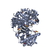
| Deposited unit | 
| ||||||||
|---|---|---|---|---|---|---|---|---|---|
| 1 |
| ||||||||
| Unit cell |
|
-Components
| #1: Protein | Mass: 46451.355 Da / Num. of mol.: 1 / Fragment: UNP residues 26-412 / Mutation: M144D Source method: isolated from a genetically manipulated source Source: (gene. exp.) Pasteurella multocida (bacteria) / Strain: P-1059 / Gene: homologue of Pm0188 / Plasmid: pET23a(+) / Production host: References: UniProt: Q15KI8, Transferases; Glycosyltransferases; Transferring other glycosyl groups |
|---|---|
| #2: Chemical | ChemComp-FN5 / 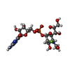  Mass: 588.432 Da / Num. of mol.: 1 / Source method: obtained synthetically / Formula: C19H30FN4O14P Mass: 588.432 Da / Num. of mol.: 1 / Source method: obtained synthetically / Formula: C19H30FN4O14P |
| #3: Water | ChemComp-HOH /  Mass: 18.015 Da / Num. of mol.: 431 / Source method: isolated from a natural source / Formula: H2O Mass: 18.015 Da / Num. of mol.: 431 / Source method: isolated from a natural source / Formula: H2O |
-Experimental details
-Experiment
| Experiment | Method: X-RAY DIFFRACTION / Number of used crystals: 1 |
|---|
- Sample preparation
| Crystal | Density Matthews: 1.99 Å3/Da / Density % sol: 38.23 % |
|---|---|
| Crystal grow | Temperature: 293 K / Method: vapor diffusion, hanging drop / pH: 7.5 Details: 24% PEG3350, 50 mM sodium chloride, 0.4% Triton X-100, 100 mM HEPES , pH 7.5, VAPOR DIFFUSION, HANGING DROP, temperature 293K |
-Data collection
| Diffraction | Mean temperature: 100 K |
|---|---|
| Diffraction source | Source: SYNCHROTRON / Site: SSRL  / Beamline: BL9-2 / Wavelength: 0.97946 / Beamline: BL9-2 / Wavelength: 0.97946 |
| Detector | Type: MARMOSAIC 325 mm CCD / Detector: CCD / Date: Nov 24, 2010 |
| Radiation | Monochromator: Si 111 Double crystal monochromator / Protocol: SINGLE WAVELENGTH / Monochromatic (M) / Laue (L): M / Scattering type: x-ray |
| Radiation wavelength | Wavelength: 0.97946 Å / Relative weight: 1 |
| Reflection | Resolution: 1.45→25 Å / Num. all: 64577 / Num. obs: 63327 / % possible obs: 98.1 % / Observed criterion σ(F): 0 / Observed criterion σ(I): -3 / Redundancy: 3.6 % / Rmerge(I) obs: 0.034 / Rsym value: 0.034 / Net I/σ(I): 19.06 |
| Reflection shell | Resolution: 1.45→1.49 Å / Redundancy: 3.3 % / Rmerge(I) obs: 0.455 / Mean I/σ(I) obs: 2.58 / Num. unique all: 5052 / % possible all: 98.2 |
- Processing
| Software |
| ||||||||||||||||||||||||||||||||||||||||||||||||||||||||||||||||||||||||||||||||||||||||||||||||||||||||||||||||||||||||||||||||||||||||||||||||||||||||||||||||||||||||||
|---|---|---|---|---|---|---|---|---|---|---|---|---|---|---|---|---|---|---|---|---|---|---|---|---|---|---|---|---|---|---|---|---|---|---|---|---|---|---|---|---|---|---|---|---|---|---|---|---|---|---|---|---|---|---|---|---|---|---|---|---|---|---|---|---|---|---|---|---|---|---|---|---|---|---|---|---|---|---|---|---|---|---|---|---|---|---|---|---|---|---|---|---|---|---|---|---|---|---|---|---|---|---|---|---|---|---|---|---|---|---|---|---|---|---|---|---|---|---|---|---|---|---|---|---|---|---|---|---|---|---|---|---|---|---|---|---|---|---|---|---|---|---|---|---|---|---|---|---|---|---|---|---|---|---|---|---|---|---|---|---|---|---|---|---|---|---|---|---|---|---|---|
| Refinement | Method to determine structure: MOLECULAR REPLACEMENT Starting model: PDB ENTRY 2IHJ Resolution: 1.45→23.93 Å / Cor.coef. Fo:Fc: 0.964 / Cor.coef. Fo:Fc free: 0.953 / SU B: 2.995 / SU ML: 0.053 / Cross valid method: THROUGHOUT / σ(F): 0 / σ(I): 0 / ESU R: 0.079 / ESU R Free: 0.079 / Stereochemistry target values: MAXIMUM LIKELIHOOD Details: HYDROGENS HAVE BEEN ADDED IN THE RIDING POSITIONS U VALUES : RESIDUAL ONLY
| ||||||||||||||||||||||||||||||||||||||||||||||||||||||||||||||||||||||||||||||||||||||||||||||||||||||||||||||||||||||||||||||||||||||||||||||||||||||||||||||||||||||||||
| Solvent computation | Ion probe radii: 0.8 Å / Shrinkage radii: 0.8 Å / VDW probe radii: 1.4 Å / Solvent model: MASK | ||||||||||||||||||||||||||||||||||||||||||||||||||||||||||||||||||||||||||||||||||||||||||||||||||||||||||||||||||||||||||||||||||||||||||||||||||||||||||||||||||||||||||
| Displacement parameters | Biso mean: 25.652 Å2
| ||||||||||||||||||||||||||||||||||||||||||||||||||||||||||||||||||||||||||||||||||||||||||||||||||||||||||||||||||||||||||||||||||||||||||||||||||||||||||||||||||||||||||
| Refine analyze |
| ||||||||||||||||||||||||||||||||||||||||||||||||||||||||||||||||||||||||||||||||||||||||||||||||||||||||||||||||||||||||||||||||||||||||||||||||||||||||||||||||||||||||||
| Refinement step | Cycle: LAST / Resolution: 1.45→23.93 Å
| ||||||||||||||||||||||||||||||||||||||||||||||||||||||||||||||||||||||||||||||||||||||||||||||||||||||||||||||||||||||||||||||||||||||||||||||||||||||||||||||||||||||||||
| Refine LS restraints |
| ||||||||||||||||||||||||||||||||||||||||||||||||||||||||||||||||||||||||||||||||||||||||||||||||||||||||||||||||||||||||||||||||||||||||||||||||||||||||||||||||||||||||||
| LS refinement shell | Resolution: 1.45→1.488 Å / Total num. of bins used: 20
| ||||||||||||||||||||||||||||||||||||||||||||||||||||||||||||||||||||||||||||||||||||||||||||||||||||||||||||||||||||||||||||||||||||||||||||||||||||||||||||||||||||||||||
| Refinement TLS params. | Method: refined / Refine-ID: X-RAY DIFFRACTION
| ||||||||||||||||||||||||||||||||||||||||||||||||||||||||||||||||||||||||||||||||||||||||||||||||||||||||||||||||||||||||||||||||||||||||||||||||||||||||||||||||||||||||||
| Refinement TLS group |
|
