 Movie
Movie Controller


+ Open data

- Basic information
| Entry | Database: PDB / ID: 3s3z | ||||||
|---|---|---|---|---|---|---|---|
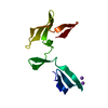
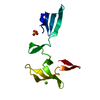
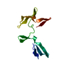
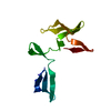
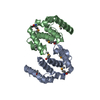
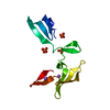
| Title | Crystal Structure an Tandem Cyanovirin-N Dimer, CVN2L10 | ||||||
 Components Components | Tandem Cyanovirin-N Dimer CVN2L10 | ||||||
 Keywords Keywords | ANTIVIRAL PROTEIN / cyanovirin-N / sugar-binding / gp120 / engineered dimer | ||||||
| Function / homology |  Function and homology information Function and homology informationoligosaccharide binding / regulation of defense response to virus / carbohydrate binding Similarity search - Function | ||||||
| Biological species |  Nostoc ellipsosporum (bacteria) Nostoc ellipsosporum (bacteria) | ||||||
| Method |  X-RAY DIFFRACTION / SYNCHROTRON / MOLECULAR REPLACEMENT / molecular replacement / Resolution: 1.75 Å X-RAY DIFFRACTION / SYNCHROTRON / MOLECULAR REPLACEMENT / molecular replacement / Resolution: 1.75 Å | ||||||
 Authors Authors | Keeffe, J.R. / Bjorkman, P.J. / Mayo, S.L. | ||||||
 Citation Citation | Journal: Proc.Natl.Acad.Sci.USA / Year: 2011 Title: Designed oligomers of cyanovirin-N show enhanced HIV neutralization. Authors: Keeffe, J.R. / Gnanapragasam, P.N. / Gillespie, S.K. / Yong, J. / Bjorkman, P.J. / Mayo, S.L. | ||||||
| History |
|
- Structure visualization
| Structure viewer | Molecule: MolmilJmol/JSmol |
|---|
- Downloads & links
-Download
| PDBx/mmCIF format | 3s3z.cif.gz | 39.4 KB | Display | PDBx/mmCIF format |
|---|---|---|---|---|
| PDB format | pdb3s3z.ent.gz | 24.9 KB | Display | PDB format |
| PDBx/mmJSON format | 3s3z.json.gz | Tree view | PDBx/mmJSON format | |
| Others |  Other downloads Other downloads |
-Validation report
| Arichive directory | https://data.pdbj.org/pub/pdb/validation_reports/s3/3s3zftp://data.pdbj.org/pub/pdb/validation_reports/s3/3s3z | HTTPS FTP |
|---|
-Related structure data
| Related structure data |  3s3yC  3ezmS C: citing same article ( S: Starting model for refinement |
|---|---|
| Similar structure data |
-Links
 PDBj
PDBj- Assembly
| Deposited unit | 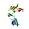
| ||||||||
|---|---|---|---|---|---|---|---|---|---|
| 1 |
| ||||||||
| Unit cell |
| ||||||||
| Details | Authors state that the biologically relevant molecule is generated by applying the crystallographic symmetry operations Y, X, -Z to the first half of the protein. |
-Components
| #1: Protein | Mass: 23999.215 Da / Num. of mol.: 1 Source method: isolated from a genetically manipulated source Source: (gene. exp.) Nostoc ellipsosporum (bacteria) / Plasmid: pET11a / Production host: | ||||
|---|---|---|---|---|---|
| #2: Chemical |   Mass: 22.990 Da / Num. of mol.: 2 / Source method: obtained synthetically / Formula: Na Mass: 22.990 Da / Num. of mol.: 2 / Source method: obtained synthetically / Formula: Na#3: Water | ChemComp-HOH / |  Mass: 18.015 Da / Num. of mol.: 87 / Source method: isolated from a natural source / Formula: H2O Mass: 18.015 Da / Num. of mol.: 87 / Source method: isolated from a natural source / Formula: H2OHas protein modification | Y | |
-Experimental details
-Experiment
| Experiment | Method: X-RAY DIFFRACTION / Number of used crystals: 1 |
|---|
- Sample preparation
| Crystal grow | Temperature: 298 K / Method: vapor diffusion, sitting drop / pH: 7.3 Details: 20% PEG 3350, 0.2 M sodium fluoride, pH 7.3, VAPOR DIFFUSION, SITTING DROP, temperature 298K |
|---|
-Data collection
| Diffraction | Mean temperature: 200 K | ||||||||||||||||||||||||||||||||||||||||||||||||||||||||||||||||||||||||||||||||||||||||
|---|---|---|---|---|---|---|---|---|---|---|---|---|---|---|---|---|---|---|---|---|---|---|---|---|---|---|---|---|---|---|---|---|---|---|---|---|---|---|---|---|---|---|---|---|---|---|---|---|---|---|---|---|---|---|---|---|---|---|---|---|---|---|---|---|---|---|---|---|---|---|---|---|---|---|---|---|---|---|---|---|---|---|---|---|---|---|---|---|---|
| Diffraction source | Source: SYNCHROTRON / Site: SSRL  / Beamline: BL12-2 / Wavelength: 0.99984 Å / Beamline: BL12-2 / Wavelength: 0.99984 Å | ||||||||||||||||||||||||||||||||||||||||||||||||||||||||||||||||||||||||||||||||||||||||
| Detector | Type: MARMOSAIC 325 mm CCD / Detector: CCD / Date: May 15, 2008 | ||||||||||||||||||||||||||||||||||||||||||||||||||||||||||||||||||||||||||||||||||||||||
| Radiation | Monochromator: Liquid nitrogen-cooled double crystal / Protocol: SINGLE WAVELENGTH / Monochromatic (M) / Laue (L): M / Scattering type: x-ray | ||||||||||||||||||||||||||||||||||||||||||||||||||||||||||||||||||||||||||||||||||||||||
| Radiation wavelength | Wavelength: 0.99984 Å / Relative weight: 1 | ||||||||||||||||||||||||||||||||||||||||||||||||||||||||||||||||||||||||||||||||||||||||
| Reflection | Resolution: 1.75→41.56 Å / Num. all: 11158 / Num. obs: 11158 / % possible obs: 100 % / Observed criterion σ(F): 0 / Observed criterion σ(I): 0 / Redundancy: 5.3 % / Rmerge(I) obs: 0.103 / Rsym value: 0.103 / Net I/σ(I): 12.7 | ||||||||||||||||||||||||||||||||||||||||||||||||||||||||||||||||||||||||||||||||||||||||
| Reflection shell | Diffraction-ID: 1
|
-Phasing
| Phasing | Method: molecular replacement | |||||||||
|---|---|---|---|---|---|---|---|---|---|---|
| Phasing MR | Model details: Phaser MODE: MR_AUTO
|
- Processing
| Software |
| |||||||||||||||||||||||||||||||||||||||||||||||||||||||||||||||||
|---|---|---|---|---|---|---|---|---|---|---|---|---|---|---|---|---|---|---|---|---|---|---|---|---|---|---|---|---|---|---|---|---|---|---|---|---|---|---|---|---|---|---|---|---|---|---|---|---|---|---|---|---|---|---|---|---|---|---|---|---|---|---|---|---|---|---|
| Refinement | Method to determine structure: MOLECULAR REPLACEMENT Starting model: PDB entry 3EZM Resolution: 1.75→36.81 Å / Cor.coef. Fo:Fc: 0.951 / Cor.coef. Fo:Fc free: 0.945 / WRfactor Rfree: 0.215 / WRfactor Rwork: 0.1993 / Occupancy max: 1 / Occupancy min: 0 / FOM work R set: 0.875 / SU B: 2.26 / SU ML: 0.074 / SU R Cruickshank DPI: 0.1209 / SU Rfree: 0.1102 / Cross valid method: THROUGHOUT / σ(F): 0 / ESU R Free: 0.11 / Stereochemistry target values: MAXIMUM LIKELIHOOD Details: (1) Only half of the crystallized molecule is in the asymmetric unit. The entire polypeptide chain (biologically relevant molecule) is generated by the operations: Y, X, -Z. (2) Because only ...Details: (1) Only half of the crystallized molecule is in the asymmetric unit. The entire polypeptide chain (biologically relevant molecule) is generated by the operations: Y, X, -Z. (2) Because only half of the biologically relevant molecule is in the asymmetric unit, the density corresponding to residue 0 in the structure is from both an Arg in the expression tag (residue 0 of the sequence) and a Gly from the linker (residue 111 of the sequence). Since side chain density was not seen, the backbone was modeled at 100% occupancy and the side chain was not modeled. (3) HYDROGENS HAVE BEEN ADDED IN THE RIDING POSITIONS.
| |||||||||||||||||||||||||||||||||||||||||||||||||||||||||||||||||
| Solvent computation | Ion probe radii: 0.8 Å / Shrinkage radii: 0.8 Å / VDW probe radii: 1.2 Å / Solvent model: MASK | |||||||||||||||||||||||||||||||||||||||||||||||||||||||||||||||||
| Displacement parameters | Biso max: 48.95 Å2 / Biso mean: 20.3458 Å2 / Biso min: 10.66 Å2
| |||||||||||||||||||||||||||||||||||||||||||||||||||||||||||||||||
| Refinement step | Cycle: LAST / Resolution: 1.75→36.81 Å
| |||||||||||||||||||||||||||||||||||||||||||||||||||||||||||||||||
| Refine LS restraints |
| |||||||||||||||||||||||||||||||||||||||||||||||||||||||||||||||||
| LS refinement shell | Resolution: 1.75→1.796 Å / Total num. of bins used: 20
|
