- PDB-3s2v: Crystal Structure of the Ligand Binding Domain of GluK1 in Comple... -
+
Open data
ID or keywords:
Loading...
-
Basic information
Entry
Database: PDB / ID: 3s2v
Title
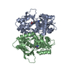
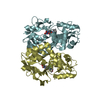
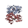
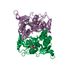
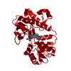
Crystal Structure of the Ligand Binding Domain of GluK1 in Complex with an Antagonist (S)-1-(2'-Amino-2'-carboxyethyl)-3-[(2-carboxythien-3-yl)methyl]thieno[3,4-d]pyrimidin-2,4-dione at 2.5 A Resolution
Components
Glutamate receptor, ionotropic kainate 1
Keywords
MEMBRANE PROTEIN / antagonist / Ionotropic Glutamate Receptor
Function / homology
Function and homology information
negative regulation of synaptic transmission, GABAergic / L-glutamate transmembrane transporter activity / positive regulation of gamma-aminobutyric acid secretion / Activation of Na-permeable kainate receptors / Activation of Ca-permeable Kainate Receptor / kainate selective glutamate receptor complex / regulation of short-term neuronal synaptic plasticity / negative regulation of synaptic transmission, glutamatergic / glutamate binding / inhibitory postsynaptic potential ...negative regulation of synaptic transmission, GABAergic / L-glutamate transmembrane transporter activity / positive regulation of gamma-aminobutyric acid secretion / Activation of Na-permeable kainate receptors / Activation of Ca-permeable Kainate Receptor / kainate selective glutamate receptor complex / regulation of short-term neuronal synaptic plasticity / negative regulation of synaptic transmission, glutamatergic / glutamate binding / inhibitory postsynaptic potential / adult behavior / kainate selective glutamate receptor activity / behavioral response to pain / extracellularly glutamate-gated ion channel activity / modulation of excitatory postsynaptic potential / ionotropic glutamate receptor complex / membrane depolarization / glutamate-gated receptor activity / glutamate-gated calcium ion channel activity / ionotropic glutamate receptor signaling pathway / establishment of localization in cell / ligand-gated monoatomic ion channel activity involved in regulation of presynaptic membrane potential / presynaptic modulation of chemical synaptic transmission / positive regulation of synaptic transmission, GABAergic / SNARE binding / transmitter-gated monoatomic ion channel activity involved in regulation of postsynaptic membrane potential / synaptic transmission, glutamatergic / regulation of membrane potential / excitatory postsynaptic potential / regulation of synaptic plasticity / postsynaptic density membrane / modulation of chemical synaptic transmission / terminal bouton / nervous system development / scaffold protein binding / presynaptic membrane / chemical synaptic transmission / postsynaptic membrane / signaling receptor complex / postsynaptic density / neuronal cell body / synapse / dendrite / glutamatergic synapse / membrane / identical protein binding / plasma membrane Similarity search - Function
Ionotropic glutamate receptor, metazoa / Ligated ion channel L-glutamate- and glycine-binding site / Ligand-gated ion channel / Ionotropic glutamate receptor, L-glutamate and glycine-binding domain / Ligated ion channel L-glutamate- and glycine-binding site / : / Ionotropic glutamate receptor / Eukaryotic homologues of bacterial periplasmic substrate binding proteins. / Periplasmic binding protein-like II / Receptor, ligand binding region ...Ionotropic glutamate receptor, metazoa / Ligated ion channel L-glutamate- and glycine-binding site / Ligand-gated ion channel / Ionotropic glutamate receptor, L-glutamate and glycine-binding domain / Ligated ion channel L-glutamate- and glycine-binding site / : / Ionotropic glutamate receptor / Eukaryotic homologues of bacterial periplasmic substrate binding proteins. / Periplasmic binding protein-like II / Receptor, ligand binding region / Receptor family ligand binding region / D-Maltodextrin-Binding Protein; domain 2 / Periplasmic binding protein-like I / 3-Layer(aba) Sandwich / Alpha Beta Similarity search - Domain/homology
Mass: 18.015 Da / Num. of mol.: 330 / Source method: isolated from a natural source / Formula: H2O
-
Details
Has protein modification
Y
Sequence details
THE STRUCTURE IS REPRESENTATIVE OF SEGMENTS S1 AND S2 COMPRISING THE LIGAND BINDING DOMAIN OF THE ...THE STRUCTURE IS REPRESENTATIVE OF SEGMENTS S1 AND S2 COMPRISING THE LIGAND BINDING DOMAIN OF THE FULL LENGTH GLUK1, CONNECTED WITH A GT LINKER. THEREFORE, SEQRES MATCHES DISCONTINUOUSLY WITH REFERENCE DATABASE (430-544, 667-805 OF ISOFORM GLUR5-2). DBREF RECORDS REFERS TO UNP ENTRY P22756-2 .
-
Experimental details
-
Experiment
Experiment
Method: X-RAY DIFFRACTION / Number of used crystals: 1
-
Sample preparation
Crystal
Density Matthews: 2.64 Å3/Da / Density % sol: 53.42 %
Crystal grow
Temperature: 279 K / Method: vapor diffusion, hanging drop / pH: 4.5 Details: 24.4% PEG4000, 0.3 M lithium sulfate, 0.1 M phosphate-citrate, pH 4.5, VAPOR DIFFUSION, HANGING DROP, temperature 279K
-
Data collection
Diffraction
Mean temperature: 100 K
Diffraction source
Source: SYNCHROTRON / Site: MAX II / Beamline: I911-3 / Wavelength: 1 Å
Detector
Type: MARMOSAIC 225 mm CCD / Detector: CCD / Date: Oct 21, 2009
Radiation
Protocol: SINGLE WAVELENGTH / Monochromatic (M) / Laue (L): M / Scattering type: x-ray
Radiation wavelength
Wavelength: 1 Å / Relative weight: 1
Reflection
Resolution: 2.5→29.3 Å / Num. obs: 21923 / % possible obs: 99.8 % / Redundancy: 3.7 % / Rmerge(I) obs: 0.094 / Net I/σ(I): 5.6
Reflection shell
Resolution: 2.5→2.64 Å / Redundancy: 3.7 % / Rmerge(I) obs: 0.24 / Mean I/σ(I) obs: 1.8 / % possible all: 100
In the structure databanks used in Yorodumi, some data are registered as the other names, "COVID-19 virus" and "2019-nCoV". Here are the details of the virus and the list of structure data.
Jan 31, 2019. EMDB accession codes are about to change! (news from PDBe EMDB page)
EMDB accession codes are about to change! (news from PDBe EMDB page)
The allocation of 4 digits for EMDB accession codes will soon come to an end. Whilst these codes will remain in use, new EMDB accession codes will include an additional digit and will expand incrementally as the available range of codes is exhausted. The current 4-digit format prefixed with “EMD-” (i.e. EMD-XXXX) will advance to a 5-digit format (i.e. EMD-XXXXX), and so on. It is currently estimated that the 4-digit codes will be depleted around Spring 2019, at which point the 5-digit format will come into force.
The EM Navigator/Yorodumi systems omit the EMD- prefix.
Related info.:Q: What is EMD? / ID/Accession-code notation in Yorodumi/EM Navigator
Yorodumi is a browser for structure data from EMDB, PDB, SASBDB, etc.
This page is also the successor to EM Navigator detail page, and also detail information page/front-end page for Omokage search.
The word "yorodu" (or yorozu) is an old Japanese word meaning "ten thousand". "mi" (miru) is to see.
Related info.:EMDB / PDB / SASBDB / Comparison of 3 databanks / Yorodumi Search / Aug 31, 2016. New EM Navigator & Yorodumi / Yorodumi Papers / Jmol/JSmol / Function and homology information / Changes in new EM Navigator and Yorodumi
 Movie
Movie Controller

 Yorodumi
 Open data
 Basic information
 Components
 Keywords
 Function and homology information
 X-RAY DIFFRACTION /
  Authors
 Citation
 Structure visualization
 Downloads & links
 Other downloads


 PDBj
PDBj

 Assembly


 Mass: 395.410 Da / Num. of mol.: 2 / Source method: obtained synthetically / Formula: C15H13N3O6S2
 Mass: 96.063 Da / Num. of mol.: 3 / Source method: obtained synthetically / Formula: SO4
 Mass: 92.094 Da / Num. of mol.: 2 / Source method: obtained synthetically / Formula: C3H8O3
 Mass: 35.453 Da / Num. of mol.: 1 / Source method: obtained synthetically / Formula: Cl
 Sample preparation
 / Beamline: I911-3 / Wavelength: 1 Å
 Processing