- PDB-3rzc: Structure of the self-antigen iGb3 bound to mouse CD1d and in com... -
+
Open data
ID or keywords:
Loading...
-
Basic information
Entry
Database: PDB / ID: 3rzc
Title
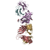
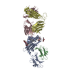
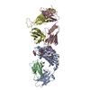
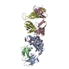
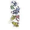
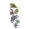
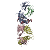
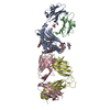
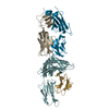
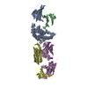
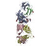
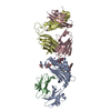
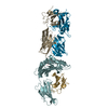
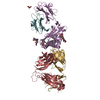
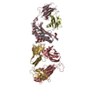
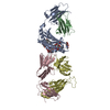
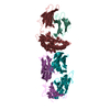
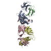
Structure of the self-antigen iGb3 bound to mouse CD1d and in complex with the iNKT TCR
Components
Antigen-presenting glycoprotein CD1d1
Beta-2-microglobulin
Valpha14
Vbeta8.2
Keywords
IMMUNE SYSTEM / antigen presentation / glycolipid / NKT cells
Function / homology
Function and homology information
regulation of immature T cell proliferation in thymus / lipid antigen binding / positive regulation of NK T cell activation / positive regulation of NK T cell differentiation / NK T cell differentiation / endogenous lipid antigen binding / exogenous lipid antigen binding / antigen processing and presentation, endogenous lipid antigen via MHC class Ib / lipopeptide binding / antigen processing and presentation, exogenous lipid antigen via MHC class Ib ...regulation of immature T cell proliferation in thymus / lipid antigen binding / positive regulation of NK T cell activation / positive regulation of NK T cell differentiation / NK T cell differentiation / endogenous lipid antigen binding / exogenous lipid antigen binding / antigen processing and presentation, endogenous lipid antigen via MHC class Ib / lipopeptide binding / antigen processing and presentation, exogenous lipid antigen via MHC class Ib / positive thymic T cell selection / positive regulation of macrophage activation / Endosomal/Vacuolar pathway / DAP12 interactions / Antigen Presentation: Folding, assembly and peptide loading of class I MHC / ER-Phagosome pathway / DAP12 signaling / Immunoregulatory interactions between a Lymphoid and a non-Lymphoid cell / antigen processing and presentation / regulation of membrane depolarization / positive regulation of interleukin-4 production / regulation of immune response / positive regulation of interleukin-2 production / cellular defense response / T cell receptor binding / Neutrophil degranulation / positive regulation of T cell proliferation / cell adhesion molecule binding / regulation of iron ion transport / cellular response to iron(III) ion / negative regulation of iron ion transport / negative regulation of forebrain neuron differentiation / antigen processing and presentation of exogenous protein antigen via MHC class Ib, TAP-dependent / iron ion transport / peptide antigen assembly with MHC class I protein complex / regulation of erythrocyte differentiation / response to molecule of bacterial origin / HFE-transferrin receptor complex / MHC class I peptide loading complex / transferrin transport / cellular response to iron ion / negative regulation of receptor-mediated endocytosis / positive regulation of T cell cytokine production / antigen processing and presentation of endogenous peptide antigen via MHC class I / MHC class I protein complex / peptide antigen assembly with MHC class II protein complex / negative regulation of neurogenesis / MHC class II protein complex / cellular response to nicotine / positive regulation of receptor-mediated endocytosis / multicellular organismal-level iron ion homeostasis / positive regulation of T cell mediated cytotoxicity / antigen processing and presentation of exogenous peptide antigen via MHC class II / positive regulation of immune response / peptide antigen binding / positive regulation of type II interferon production / phagocytic vesicle membrane / positive regulation of T cell activation / negative regulation of epithelial cell proliferation / sensory perception of smell / positive regulation of cellular senescence / MHC class II protein complex binding / T cell differentiation in thymus / late endosome / late endosome membrane / antimicrobial humoral immune response mediated by antimicrobial peptide / negative regulation of neuron projection development / antibacterial humoral response / protein refolding / cellular response to lipopolysaccharide / amyloid fibril formation / protein homotetramerization / defense response to Gram-negative bacterium / intracellular iron ion homeostasis / learning or memory / early endosome / lysosome / endosome membrane / defense response to Gram-positive bacterium / immune response / external side of plasma membrane / innate immune response / lysosomal membrane / structural molecule activity / cell surface / Golgi apparatus / endoplasmic reticulum / protein homodimerization activity / : / identical protein binding / plasma membrane / cytoplasm / cytosol Similarity search - Function
MHC-I family domain / MHC class I-like antigen recognition-like / Murine Class I Major Histocompatibility Complex, H2-DB; Chain A, domain 1 / Beta-2-Microglobulin / : / MHC class I-like antigen recognition-like / MHC class I-like antigen recognition-like superfamily / MHC classes I/II-like antigen recognition protein / : / Immunoglobulin/major histocompatibility complex, conserved site ...MHC-I family domain / MHC class I-like antigen recognition-like / Murine Class I Major Histocompatibility Complex, H2-DB; Chain A, domain 1 / Beta-2-Microglobulin / : / MHC class I-like antigen recognition-like / MHC class I-like antigen recognition-like superfamily / MHC classes I/II-like antigen recognition protein / : / Immunoglobulin/major histocompatibility complex, conserved site / Immunoglobulins and major histocompatibility complex proteins signature. / Immunoglobulin C-Type / Immunoglobulin C1-set / Immunoglobulin C1-set domain / Ig-like domain profile. / Immunoglobulin-like domain / Immunoglobulin-like domain superfamily / Immunoglobulin-like fold / Immunoglobulins / Immunoglobulin-like / Sandwich / 2-Layer Sandwich / Mainly Beta / Alpha Beta Similarity search - Domain/homology
Resolution: 2.8→2.87 Å / Redundancy: 4 % / Rmerge(I) obs: 0.695 / Mean I/σ(I) obs: 2.2 / Num. unique all: 1894 / % possible all: 99.9
-
Processing
Software
Name
Version
Classification
HKL-2000
datacollection
MOLREP
phasing
REFMAC
5.5.0109
refinement
HKL-2000
datareduction
HKL-2000
datascaling
Refinement
Method to determine structure: MOLECULAR REPLACEMENT / Resolution: 2.8→49.65 Å / Cor.coef. Fo:Fc: 0.901 / Cor.coef. Fo:Fc free: 0.868 / SU B: 34.034 / SU ML: 0.3 / Cross valid method: THROUGHOUT / ESU R Free: 0.383 / Stereochemistry target values: MAXIMUM LIKELIHOOD / Details: HYDROGENS HAVE BEEN ADDED IN THE RIDING POSITIONS
Rfactor
Num. reflection
% reflection
Selection details
Rfree
0.27054
1438
5.1 %
RANDOM
Rwork
0.23555
-
-
-
obs
0.23729
26949
98.87 %
-
Solvent computation
Ion probe radii: 0.8 Å / Shrinkage radii: 0.8 Å / VDW probe radii: 1.4 Å / Solvent model: MASK
Displacement parameters
Biso mean: 46.528 Å2
Baniso -1
Baniso -2
Baniso -3
1-
-0.05 Å2
0 Å2
0 Å2
2-
-
0.09 Å2
0 Å2
3-
-
-
-0.03 Å2
Refinement step
Cycle: LAST / Resolution: 2.8→49.65 Å
Protein
Nucleic acid
Ligand
Solvent
Total
Num. atoms
6360
0
186
48
6594
Refine LS restraints
Refine-ID
Type
Dev ideal
Dev ideal target
Number
X-RAY DIFFRACTION
r_bond_refined_d
0.005
0.022
6731
X-RAY DIFFRACTION
r_angle_refined_deg
0.835
1.964
9168
X-RAY DIFFRACTION
r_dihedral_angle_1_deg
4.144
5
801
X-RAY DIFFRACTION
r_dihedral_angle_2_deg
33.344
24.369
309
X-RAY DIFFRACTION
r_dihedral_angle_3_deg
12.049
15
1037
X-RAY DIFFRACTION
r_dihedral_angle_4_deg
11.812
15
34
X-RAY DIFFRACTION
r_chiral_restr
0.047
0.2
1017
X-RAY DIFFRACTION
r_gen_planes_refined
0.002
0.021
5098
X-RAY DIFFRACTION
r_mcbond_it
0.076
1.5
4025
X-RAY DIFFRACTION
r_mcangle_it
0.145
2
6508
X-RAY DIFFRACTION
r_scbond_it
0.184
3
2706
X-RAY DIFFRACTION
r_scangle_it
0.341
4.5
2660
LS refinement shell
Resolution: 2.798→2.871 Å / Total num. of bins used: 20
Rfactor
Num. reflection
% reflection
Rfree
0.392
111
-
Rwork
0.323
1934
-
obs
-
-
97.89 %
Refinement TLS params.
Method: refined / Refine-ID: X-RAY DIFFRACTION
ID
L11 (°2)
L12 (°2)
L13 (°2)
L22 (°2)
L23 (°2)
L33 (°2)
S11 (Å °)
S12 (Å °)
S13 (Å °)
S21 (Å °)
S22 (Å °)
S23 (Å °)
S31 (Å °)
S32 (Å °)
S33 (Å °)
T11 (Å2)
T12 (Å2)
T13 (Å2)
T22 (Å2)
T23 (Å2)
T33 (Å2)
Origin x (Å)
Origin y (Å)
Origin z (Å)
1
2.4722
-1.4101
-0.5024
2.9262
1.3964
4.2087
-0.0461
0.0184
-0.0053
-0.031
0.0007
0.1449
0.0514
-0.0945
0.0454
0.0197
-0.0185
-0.0286
0.0559
0.0295
0.0474
33.6074
20.8323
-17.7478
2
7.0487
-0.1312
0.7233
2.9235
-0.2198
3.3273
0.3147
-0.7131
-0.6279
0.3745
-0.2505
-0.662
0.2955
0.4434
-0.0642
0.186
-0.0171
0.0374
0.1809
0.1107
0.492
64.4001
-0.8582
-16.7921
3
8.7085
-0.5139
-4.1006
3.6343
1.4144
3.4387
-0.0959
-0.4092
0.2307
-0.1827
0.0898
-0.5987
-0.142
0.3432
0.0061
0.117
-0.0502
0.002
0.1531
0.0259
0.2214
63.0148
20.0272
-20.783
4
5.0349
-1.7741
1.8951
0.9785
-0.2333
3.0372
0.0537
0.1159
-0.2067
-0.0911
0.0005
0.0546
-0.1371
-0.0381
-0.0542
0.0892
-0.011
-0.0227
0.0212
-0.0165
0.0464
6.3653
39.1936
-26.3018
5
6.4561
1.2238
0.669
8.6303
1.646
5.6334
-0.2799
0.7351
0.2982
-1.0361
0.0837
0.5932
-0.5718
0.0618
0.1963
0.2705
0.1176
-0.1311
0.3994
0.0362
0.1021
-16.618
67.5916
-28.7389
6
1.2133
-1.6967
0.2644
5.9149
-0.4527
1.0049
-0.0422
-0.1386
0.2216
0.3606
0.0355
-0.2871
-0.0931
-0.1321
0.0066
0.1039
-0.0342
-0.0198
0.0785
-0.0381
0.0691
18.5257
53.0797
-12.0507
7
8.4327
2.4589
-0.0702
3.6222
0.1149
2.5124
-0.0091
0.3277
0.1654
-0.1659
0.0253
-0.0305
-0.1839
-0.0308
-0.0161
0.1242
0.0516
-0.0117
0.0366
-0.004
0.0299
-6.3353
70.0873
-14.6926
Refinement TLS group
ID
Refine-ID
Refine TLS-ID
Auth asym-ID
Auth seq-ID
1
X-RAY DIFFRACTION
1
A
6 - 180
2
X-RAY DIFFRACTION
1
A
500 - 507
3
X-RAY DIFFRACTION
1
A
286
4
X-RAY DIFFRACTION
2
A
181 - 279
5
X-RAY DIFFRACTION
3
B
2 - 99
6
X-RAY DIFFRACTION
4
C
1 - 112
7
X-RAY DIFFRACTION
5
C
113 - 205
8
X-RAY DIFFRACTION
6
D
2 - 112
9
X-RAY DIFFRACTION
7
D
113 - 240
+
About Yorodumi
-
News
-
Feb 9, 2022. New format data for meta-information of EMDB entries
New format data for meta-information of EMDB entries
Version 3 of the EMDB header file is now the official format.
The previous official version 1.9 will be removed from the archive.
In the structure databanks used in Yorodumi, some data are registered as the other names, "COVID-19 virus" and "2019-nCoV". Here are the details of the virus and the list of structure data.
Jan 31, 2019. EMDB accession codes are about to change! (news from PDBe EMDB page)
EMDB accession codes are about to change! (news from PDBe EMDB page)
The allocation of 4 digits for EMDB accession codes will soon come to an end. Whilst these codes will remain in use, new EMDB accession codes will include an additional digit and will expand incrementally as the available range of codes is exhausted. The current 4-digit format prefixed with “EMD-” (i.e. EMD-XXXX) will advance to a 5-digit format (i.e. EMD-XXXXX), and so on. It is currently estimated that the 4-digit codes will be depleted around Spring 2019, at which point the 5-digit format will come into force.
The EM Navigator/Yorodumi systems omit the EMD- prefix.
Related info.:Q: What is EMD? / ID/Accession-code notation in Yorodumi/EM Navigator
Yorodumi is a browser for structure data from EMDB, PDB, SASBDB, etc.
This page is also the successor to EM Navigator detail page, and also detail information page/front-end page for Omokage search.
The word "yorodu" (or yorozu) is an old Japanese word meaning "ten thousand". "mi" (miru) is to see.
Related info.:EMDB / PDB / SASBDB / Comparison of 3 databanks / Yorodumi Search / Aug 31, 2016. New EM Navigator & Yorodumi / Yorodumi Papers / Jmol/JSmol / Function and homology information / Changes in new EM Navigator and Yorodumi
 Movie
Movie Controller

 Yorodumi
 Open data
 Basic information
 Components
 Keywords
 Function and homology information
 Homo sapiens (human)
 X-RAY DIFFRACTION /
  Authors
 Citation
 Structure visualization
 Downloads & links
 Other downloads
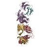

 PDBj
PDBj


 Assembly
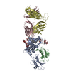

 Spodoptera frugiperda (fall armyworm) / Strain (production host): Sf9 / References: UniProt: P11609

 Mass: 1164.588 Da / Num. of mol.: 1 / Source method: obtained synthetically / Formula: C62H117NO18
 Sample preparation
 / Beamline: BL7-1 / Wavelength: 0.979 Å
 Processing