Protocol: SINGLE WAVELENGTH / Monochromatic (M) / Laue (L): M / Scattering type: x-ray
Radiation wavelength
Wavelength: 0.97942 Å / Relative weight: 1
Reflection
Redundancy: 4.4 % / Av σ(I) over netI: 23.92 / Number: 491490 / Rmerge(I) obs: 0.088 / Χ2: 1.8 / D res high: 1.5 Å / D res low: 50 Å / Num. obs: 111443 / % possible obs: 99.2
D res high: 2 Å / D res low: 50 Å / FOM : 0.387 / FOM acentric: 0.4 / FOM centric: 0 / Reflection: 47424 / Reflection acentric: 45870 / Reflection centric: 1554
Phasing MAD set
R cullis acentric: 1.42 / R cullis centric: 1 / Highest resolution: 2 Å / Lowest resolution: 50 Å / Loc acentric: 0.2 / Loc centric: 0.2 / Power acentric: 0 / Power centric: 0 / Reflection acentric: 45870 / Reflection centric: 1554
Phasing MAD set shell
ID: 1 / R cullis centric: 1 / Power acentric: 0 / Power centric: 0
Resolution (Å)
R cullis acentric
Loc acentric
Loc centric
Reflection acentric
Reflection centric
12.5-50
1.51
0.3
0.2
162
34
7.14-12.5
1.37
0.3
0.2
788
81
5-7.14
1.63
0.3
0.2
1896
127
3.85-5
1.02
0.2
0.2
3465
164
3.13-3.85
1.11
0.2
0.2
5578
221
2.63-3.13
1.49
0.2
0.1
8115
264
2.27-2.63
1.62
0.2
0.1
11183
310
2-2.27
1.56
0.2
0.1
14683
353
Phasing MAD set site
Atom type symbol: Se / Occupancy iso: 0
ID
B iso
Fract x
Fract y
Fract z
Occupancy
1
8.2291
-0.167
-0.671
-0.004
4.912
2
10.0233
-0.072
-0.713
0.327
4.561
3
8.9183
-0.391
-0.632
-0.089
3.718
4
10.7836
-0.321
-0.501
-0.105
3.924
5
8.69
0.081
-0.531
0.403
3.524
6
12.316
0.153
-0.662
0.405
3.476
7
31.8865
-0.427
-0.582
-0.219
3.825
8
14.5077
0.138
-0.696
0.261
2.96
9
50.3734
0.192
-0.597
0.527
3.835
10
17.0174
-0.348
-0.661
0.072
1.866
11
50.4192
-0.626
-0.46
-0.082
3.303
12
11.3476
0.189
-0.642
0.41
1.584
13
6.2454
-0.428
-0.613
-0.097
1.628
14
44.7017
0.382
-0.491
0.373
2.317
Phasing MAD shell
Resolution (Å)
FOM
FOM acentric
FOM centric
Reflection
Reflection acentric
Reflection centric
12.5-50
0.233
0.282
0
196
162
34
7.14-12.5
0.398
0.439
0
869
788
81
5-7.14
0.442
0.471
0
2023
1896
127
3.85-5
0.357
0.374
0
3629
3465
164
3.13-3.85
0.389
0.405
0
5799
5578
221
2.63-3.13
0.406
0.419
0
8379
8115
264
2.27-2.63
0.403
0.415
0
11493
11183
310
2-2.27
0.365
0.374
0
15036
14683
353
Phasing dm
Method: Solvent flattening and Histogram matching / Reflection: 111310
Phasing dm shell
Resolution (Å)
Delta phi final
FOM
Reflection
8.13-100
60.9
0.781
721
5.75-8.13
58.7
0.893
1314
4.69-5.75
59.9
0.915
1695
4.07-4.69
61.7
0.922
1998
3.64-4.07
62.5
0.912
2243
3.32-3.64
62.3
0.904
2469
3.07-3.32
63.7
0.884
2708
2.87-3.07
63.3
0.876
2897
2.71-2.87
62.1
0.886
3084
2.57-2.71
61.9
0.875
3264
2.45-2.57
61.9
0.881
3457
2.35-2.45
61.6
0.877
3538
2.26-2.35
61
0.873
3772
2.17-2.26
62.6
0.876
3873
2.1-2.17
64
0.871
3991
2.03-2.1
64.4
0.865
4171
1.97-2.03
76.7
0.85
4272
1.92-1.97
90.5
0.835
4403
1.87-1.92
87.1
0.825
4541
1.82-1.87
89.1
0.825
4655
1.77-1.82
90.1
0.819
4750
1.73-1.77
89.3
0.809
4912
1.7-1.73
89.9
0.8
4971
1.66-1.7
90.1
0.797
5112
1.63-1.66
90.3
0.782
5192
1.59-1.63
89.5
0.771
5295
1.56-1.59
89.2
0.733
5450
1.54-1.56
92.2
0.699
5488
1.5-1.54
89.9
0.54
7074
-
Processing
Software
Name
Version
Classification
NB
DENZO
datareduction
SCALEPACK
datascaling
MLPHARE
phasing
DM
6.1
phasing
REFMAC
refinement
PDB_EXTRACT
3.1
dataextraction
SBC-Collect
datacollection
HKL-3000
datareduction
HKL-3000
datascaling
HKL-3000
phasing
SHELXD
phasing
SHELXE
modelbuilding
SOLVE
phasing
RESOLVE
phasing
ARP/wARP
modelbuilding
CCP4
phasing
O
modelbuilding
Coot
modelbuilding
Refinement
Method to determine structure: SAD / Resolution: 1.5→36.5 Å / Cor.coef. Fo:Fc: 0.957 / Cor.coef. Fo:Fc free: 0.946 / Occupancy max: 1 / Occupancy min: 0.5 / SU B: 2.188 / SU ML: 0.041 / SU R Cruickshank DPI: 0.0716 / Cross valid method: THROUGHOUT / ESU R: 0.072 / ESU R Free: 0.072 Stereochemistry target values: MAXIMUM LIKELIHOOD WITH PHASES Details: HYDROGENS HAVE BEEN ADDED IN THE RIDING POSITIONS
Rfactor
Num. reflection
% reflection
Selection details
Rfree
0.189
5565
5 %
RANDOM
Rwork
0.16362
-
-
-
obs
0.1649
105371
99.14 %
-
all
-
110936
-
-
Solvent computation
Ion probe radii: 0.8 Å / Shrinkage radii: 0.8 Å / VDW probe radii: 1.4 Å / Solvent model: MASK
Displacement parameters
Biso mean: 12.229 Å2
Baniso -1
Baniso -2
Baniso -3
1-
-0.33 Å2
0 Å2
0.54 Å2
2-
-
-0.17 Å2
0 Å2
3-
-
-
0.55 Å2
Refinement step
Cycle: LAST / Resolution: 1.5→36.5 Å
Protein
Nucleic acid
Ligand
Solvent
Total
Num. atoms
5438
0
38
944
6420
Refine LS restraints
Refine-ID
Type
Dev ideal
Dev ideal target
Number
X-RAY DIFFRACTION
r_bond_refined_d
0.013
0.022
5668
X-RAY DIFFRACTION
r_bond_other_d
0.001
0.02
3867
X-RAY DIFFRACTION
r_angle_refined_deg
1.425
1.967
7692
X-RAY DIFFRACTION
r_angle_other_deg
0.905
3
9496
X-RAY DIFFRACTION
r_dihedral_angle_1_deg
5.326
5
706
X-RAY DIFFRACTION
r_dihedral_angle_2_deg
35.69
24.859
249
X-RAY DIFFRACTION
r_dihedral_angle_3_deg
11.735
15
1028
X-RAY DIFFRACTION
r_dihedral_angle_4_deg
15.315
15
29
X-RAY DIFFRACTION
r_chiral_restr
0.085
0.2
870
X-RAY DIFFRACTION
r_gen_planes_refined
0.006
0.021
6241
X-RAY DIFFRACTION
r_gen_planes_other
0.001
0.02
1076
X-RAY DIFFRACTION
r_nbd_refined
X-RAY DIFFRACTION
r_nbd_other
X-RAY DIFFRACTION
r_nbtor_refined
X-RAY DIFFRACTION
r_nbtor_other
X-RAY DIFFRACTION
r_xyhbond_nbd_refined
X-RAY DIFFRACTION
r_xyhbond_nbd_other
X-RAY DIFFRACTION
r_metal_ion_refined
X-RAY DIFFRACTION
r_metal_ion_other
X-RAY DIFFRACTION
r_symmetry_vdw_refined
X-RAY DIFFRACTION
r_symmetry_vdw_other
X-RAY DIFFRACTION
r_symmetry_hbond_refined
X-RAY DIFFRACTION
r_symmetry_hbond_other
X-RAY DIFFRACTION
r_symmetry_metal_ion_refined
X-RAY DIFFRACTION
r_symmetry_metal_ion_other
X-RAY DIFFRACTION
r_mcbond_it
0.762
1.5
3487
X-RAY DIFFRACTION
r_mcbond_other
0.229
1.5
1399
X-RAY DIFFRACTION
r_mcangle_it
1.361
2
5657
X-RAY DIFFRACTION
r_scbond_it
2.347
3
2181
X-RAY DIFFRACTION
r_scangle_it
3.757
4.5
2032
X-RAY DIFFRACTION
r_rigid_bond_restr
X-RAY DIFFRACTION
r_sphericity_free
X-RAY DIFFRACTION
r_sphericity_bonded
LS refinement shell
Resolution: 1.5→1.539 Å / Total num. of bins used: 20
Rfactor
Num. reflection
% reflection
Rfree
0.219
410
-
Rwork
0.198
7787
-
obs
-
-
98.93 %
Refinement TLS params.
Method: refined / Refine-ID: X-RAY DIFFRACTION
ID
L11 (°2)
L12 (°2)
L13 (°2)
L22 (°2)
L23 (°2)
L33 (°2)
S11 (Å °)
S12 (Å °)
S13 (Å °)
S21 (Å °)
S22 (Å °)
S23 (Å °)
S31 (Å °)
S32 (Å °)
S33 (Å °)
T11 (Å2)
T12 (Å2)
T13 (Å2)
T22 (Å2)
T23 (Å2)
T33 (Å2)
Origin x (Å)
Origin y (Å)
Origin z (Å)
1
0.3707
0.1799
-0.0143
0.3643
0.0281
0.2785
-0.0042
0.038
-0.0118
0.0129
-0.0047
-0.0199
-0.0039
0.0137
0.0089
0.0237
0.0098
-0.0055
0.0305
0.002
0.0032
2.1676
33.6449
35.5847
2
0.4573
-0.1286
-0.0012
0.2918
-0.0596
0.312
0.006
0.016
0.0151
-0.0253
-0.0054
0.0034
-0.0008
-0.0085
-0.0006
0.018
-0.0052
-0.0069
0.0156
-0.0011
0.0097
-18.0337
35.9092
78.1541
Refinement TLS group
ID
Refine-ID
Refine TLS-ID
Auth asym-ID
Auth seq-ID
1
X-RAY DIFFRACTION
1
A
-10 - 9999
2
X-RAY DIFFRACTION
2
B
-10 - 9999
+
About Yorodumi
-
News
-
Feb 9, 2022. New format data for meta-information of EMDB entries
New format data for meta-information of EMDB entries
Version 3 of the EMDB header file is now the official format.
The previous official version 1.9 will be removed from the archive.
In the structure databanks used in Yorodumi, some data are registered as the other names, "COVID-19 virus" and "2019-nCoV". Here are the details of the virus and the list of structure data.
Jan 31, 2019. EMDB accession codes are about to change! (news from PDBe EMDB page)
EMDB accession codes are about to change! (news from PDBe EMDB page)
The allocation of 4 digits for EMDB accession codes will soon come to an end. Whilst these codes will remain in use, new EMDB accession codes will include an additional digit and will expand incrementally as the available range of codes is exhausted. The current 4-digit format prefixed with “EMD-” (i.e. EMD-XXXX) will advance to a 5-digit format (i.e. EMD-XXXXX), and so on. It is currently estimated that the 4-digit codes will be depleted around Spring 2019, at which point the 5-digit format will come into force.
The EM Navigator/Yorodumi systems omit the EMD- prefix.
Related info.:Q: What is EMD? / ID/Accession-code notation in Yorodumi/EM Navigator
Yorodumi is a browser for structure data from EMDB, PDB, SASBDB, etc.
This page is also the successor to EM Navigator detail page, and also detail information page/front-end page for Omokage search.
The word "yorodu" (or yorozu) is an old Japanese word meaning "ten thousand". "mi" (miru) is to see.
Related info.:EMDB / PDB / SASBDB / Comparison of 3 databanks / Yorodumi Search / Aug 31, 2016. New EM Navigator & Yorodumi / Yorodumi Papers / Jmol/JSmol / Function and homology information / Changes in new EM Navigator and Yorodumi
 Movie
Movie Controller

 Yorodumi
 Open data
 Basic information
 Components
 Keywords
 Function and homology information
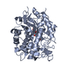
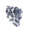
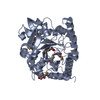
 Shewanella sp. (bacteria)
 X-RAY DIFFRACTION /
  Authors
 Citation
 Structure visualization
 Downloads & links
 Other downloads


 PDBj
PDBj Assembly
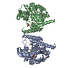

 Mass: 65.409 Da / Num. of mol.: 2 / Source method: obtained synthetically / Formula: Zn
 Type: L-peptide linking / Mass: 196.106 Da / Num. of mol.: 2 / Source method: obtained synthetically / Formula: C5H11NO2Se
 Mass: 92.094 Da / Num. of mol.: 3 / Source method: obtained synthetically / Formula: C3H8O3
 Mass: 18.015 Da / Num. of mol.: 944 / Source method: isolated from a natural source / Formula: H2O
 Sample preparation
 / Beamline: 19-ID / Wavelength: 0.97942 Å
 Processing