 Movie
Movie Controller

[English] 日本語
 Yorodumi
- PDB-3rmw: Crystal Structure of Human Glycogenin-1 (GYG1) T83M mutant comple... -
+ Open data

- Basic information
| Entry | Database: PDB / ID: 3rmw | ||||||
|---|---|---|---|---|---|---|---|
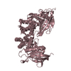
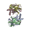
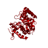
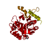
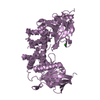
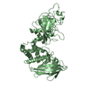
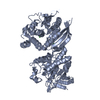
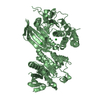
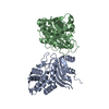
| Title | Crystal Structure of Human Glycogenin-1 (GYG1) T83M mutant complexed with manganese and UDP-glucose | ||||||
 Components Components | Glycogenin-1 | ||||||
 Keywords Keywords | TRANSFERASE / Structural Genomics / Structural Genomics Consortium / SGC / glycosyltransferase / glycogen biosynthesis / glycosylation | ||||||
| Function / homology |  Function and homology information Function and homology informationGlycogen storage disease type XV (GYG1) / Glycogen storage disease type 0 (muscle GYS1) / : / glycogenin glucosyltransferase / Glycogen storage disease type II (GAA) / glycogenin glucosyltransferase activity / glycogen biosynthetic process / Glycogen breakdown (glycogenolysis) / glycosyltransferase activity / lysosomal lumen ...Glycogen storage disease type XV (GYG1) / Glycogen storage disease type 0 (muscle GYS1) / : / glycogenin glucosyltransferase / Glycogen storage disease type II (GAA) / glycogenin glucosyltransferase activity / glycogen biosynthetic process / Glycogen breakdown (glycogenolysis) / glycosyltransferase activity / lysosomal lumen / Myoclonic epilepsy of Lafora / Glycogen synthesis / manganese ion binding / secretory granule lumen / ficolin-1-rich granule lumen / Neutrophil degranulation / protein homodimerization activity / extracellular region / membrane / nucleus / cytoplasm / cytosol Similarity search - Function | ||||||
| Biological species |  Homo sapiens (human) Homo sapiens (human) | ||||||
| Method |  X-RAY DIFFRACTION / MOLECULAR REPLACEMENT / Resolution: 1.93 Å X-RAY DIFFRACTION / MOLECULAR REPLACEMENT / Resolution: 1.93 Å | ||||||
 Authors Authors | Chaikuad, A. / Froese, D.S. / Yue, W.W. / Krysztofinska, E. / von Delft, F. / Weigelt, J. / Arrowsmith, C.H. / Edwards, A.M. / Bountra, C. / Oppermann, U. / Structural Genomics Consortium (SGC) | ||||||
 Citation Citation | Journal: Proc.Natl.Acad.Sci.USA / Year: 2011 Title: Conformational plasticity of glycogenin and its maltosaccharide substrate during glycogen biogenesis. Authors: Chaikuad, A. / Froese, D.S. / Berridge, G. / von Delft, F. / Oppermann, U. / Yue, W.W. | ||||||
| History |
|
- Structure visualization
| Structure viewer | Molecule: MolmilJmol/JSmol |
|---|
- Downloads & links
-Download
| PDBx/mmCIF format | 3rmw.cif.gz | 123.2 KB | Display | PDBx/mmCIF format |
|---|---|---|---|---|
| PDB format | pdb3rmw.ent.gz | 94.2 KB | Display | PDB format |
| PDBx/mmJSON format | 3rmw.json.gz | Tree view | PDBx/mmJSON format | |
| Others |  Other downloads Other downloads |
-Validation report
| Arichive directory | https://data.pdbj.org/pub/pdb/validation_reports/rm/3rmwftp://data.pdbj.org/pub/pdb/validation_reports/rm/3rmw | HTTPS FTP |
|---|
-Related structure data
| Related structure data |  3q4sSC 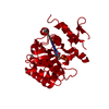 3qvbC  3rmvC  3t7mC  3t7nC 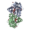 3t7oC  3u2tC  3u2uC  3u2vC  3u2wC C: citing same article ( S: Starting model for refinement |
|---|---|
| Similar structure data |

-Links
 PDBj
PDBj


- Assembly
| Deposited unit | 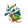
| ||||||||
|---|---|---|---|---|---|---|---|---|---|
| 1 | 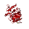
| ||||||||
| Unit cell |
|
-Components
-Protein , 1 types, 1 molecules A
| #1: Protein | Mass: 29641.729 Da / Num. of mol.: 1 / Fragment: UNP residues 1-262 / Mutation: T83M Source method: isolated from a genetically manipulated source Source: (gene. exp.) Homo sapiens (human) / Gene: GYG, GYG1 / Plasmid: pNIC28-Bsa4 / Production host:  |
|---|
-Non-polymers , 5 types, 204 molecules 


| #2: Chemical | ChemComp-UPG /  Mass: 566.302 Da / Num. of mol.: 1 / Source method: obtained synthetically / Formula: C15H24N2O17P2 Mass: 566.302 Da / Num. of mol.: 1 / Source method: obtained synthetically / Formula: C15H24N2O17P2 | ||
|---|---|---|---|
| #3: Chemical | ChemComp-MN /  Mass: 54.938 Da / Num. of mol.: 1 / Source method: obtained synthetically / Formula: Mn Mass: 54.938 Da / Num. of mol.: 1 / Source method: obtained synthetically / Formula: Mn | ||
| #4: Chemical | ChemComp-MG /  Mass: 24.305 Da / Num. of mol.: 1 / Source method: obtained synthetically / Formula: Mg Mass: 24.305 Da / Num. of mol.: 1 / Source method: obtained synthetically / Formula: Mg | ||
| #5: Chemical | ChemComp-EDO /  Mass: 62.068 Da / Num. of mol.: 12 / Source method: obtained synthetically / Formula: C2H6O2 Mass: 62.068 Da / Num. of mol.: 12 / Source method: obtained synthetically / Formula: C2H6O2#6: Water | ChemComp-HOH / | Mass: 18.015 Da / Num. of mol.: 189 / Source method: isolated from a natural source / Formula: H2O |
-Experimental details
-Experiment
| Experiment | Method: X-RAY DIFFRACTION / Number of used crystals: 1 |
|---|
- Sample preparation
| Crystal | Density Matthews: 2.36 Å3/Da / Density % sol: 47.89 % |
|---|---|
| Crystal grow | Temperature: 293.15 K / Method: vapor diffusion, sitting drop / pH: 8 Details: 25% PEG smears (PEG 6k, 8k, 10k), 0.2M MgCl2, 10% glycerol, 0.1M Tris, pH 8.0, VAPOR DIFFUSION, SITTING DROP, temperature 293.15K |
-Data collection
| Diffraction | Mean temperature: 100 K |
|---|---|
| Diffraction source | Source: ROTATING ANODE / Type: RIGAKU FR-E SUPERBRIGHT / Wavelength: 1.5418 Å |
| Detector | Type: RIGAKU RAXIS IV / Detector: IMAGE PLATE / Date: Oct 26, 2010 |
| Radiation | Monochromator: Flat graphite crystal / Protocol: SINGLE WAVELENGTH / Monochromatic (M) / Laue (L): M / Scattering type: x-ray |
| Radiation wavelength | Wavelength: 1.5418 Å / Relative weight: 1 |
| Reflection | Resolution: 1.93→29.85 Å / Num. all: 21806 / Num. obs: 21806 / % possible obs: 100 % / Observed criterion σ(F): 0 / Observed criterion σ(I): 0 / Redundancy: 4.5 % / Biso Wilson estimate: 25.5 Å2 / Rmerge(I) obs: 0.097 / Net I/σ(I): 10.7 |
| Reflection shell | Resolution: 1.93→2.03 Å / Redundancy: 4.4 % / Rmerge(I) obs: 0.728 / Mean I/σ(I) obs: 2 / Num. unique all: 3129 / % possible all: 100 |
- Processing
| Software |
| |||||||||||||||||||||||||||||||||||||||||||||||||||||||||||||||||||||||||||||||||||||||||||||||
|---|---|---|---|---|---|---|---|---|---|---|---|---|---|---|---|---|---|---|---|---|---|---|---|---|---|---|---|---|---|---|---|---|---|---|---|---|---|---|---|---|---|---|---|---|---|---|---|---|---|---|---|---|---|---|---|---|---|---|---|---|---|---|---|---|---|---|---|---|---|---|---|---|---|---|---|---|---|---|---|---|---|---|---|---|---|---|---|---|---|---|---|---|---|---|---|---|
| Refinement | Method to determine structure: MOLECULAR REPLACEMENT Starting model: PDB ENTRY 3Q4S Resolution: 1.93→28.86 Å / Cor.coef. Fo:Fc: 0.957 / Cor.coef. Fo:Fc free: 0.93 / SU B: 7.122 / SU ML: 0.109 / Cross valid method: THROUGHOUT / σ(F): 0 / σ(I): 2 / ESU R: 0.162 / ESU R Free: 0.155 / Stereochemistry target values: MAXIMUM LIKELIHOOD / Details: HYDROGENS HAVE BEEN ADDED IN THE RIDING POSITIONS
| |||||||||||||||||||||||||||||||||||||||||||||||||||||||||||||||||||||||||||||||||||||||||||||||
| Solvent computation | Ion probe radii: 0.8 Å / Shrinkage radii: 0.8 Å / VDW probe radii: 1.4 Å / Solvent model: MASK | |||||||||||||||||||||||||||||||||||||||||||||||||||||||||||||||||||||||||||||||||||||||||||||||
| Displacement parameters | Biso mean: 22.627 Å2
| |||||||||||||||||||||||||||||||||||||||||||||||||||||||||||||||||||||||||||||||||||||||||||||||
| Refine analyze | Luzzati coordinate error obs: 0.22 Å | |||||||||||||||||||||||||||||||||||||||||||||||||||||||||||||||||||||||||||||||||||||||||||||||
| Refinement step | Cycle: LAST / Resolution: 1.93→28.86 Å
| |||||||||||||||||||||||||||||||||||||||||||||||||||||||||||||||||||||||||||||||||||||||||||||||
| Refine LS restraints |
| |||||||||||||||||||||||||||||||||||||||||||||||||||||||||||||||||||||||||||||||||||||||||||||||
| LS refinement shell | Resolution: 1.93→1.98 Å / Total num. of bins used: 20
| |||||||||||||||||||||||||||||||||||||||||||||||||||||||||||||||||||||||||||||||||||||||||||||||
| Refinement TLS params. | Method: refined / Refine-ID: X-RAY DIFFRACTION
| |||||||||||||||||||||||||||||||||||||||||||||||||||||||||||||||||||||||||||||||||||||||||||||||
| Refinement TLS group |
|
