 Movie
Movie Controller

[English] 日本語
 Yorodumi
- PDB-3rmb: Crystal Structure of a replicative DNA polymerase bound to DNA co... -
+ Open data

- Basic information
| Entry | Database: PDB / ID: 3rmb | ||||||
|---|---|---|---|---|---|---|---|
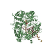
| Title | Crystal Structure of a replicative DNA polymerase bound to DNA containing Thymine Glycol | ||||||
 Components Components |
| ||||||
 Keywords Keywords | TRANSFERASE/DNA / DNA LESION / THYMINE GLYCOL / PROTEIN-DNA COMPLEX / TRANSFERASE-DNA complex | ||||||
| Function / homology |  Function and homology information Function and homology informationbidirectional double-stranded viral DNA replication / Hydrolases; Acting on ester bonds; Exodeoxyribonucleases producing 5'-phosphomonoesters / 3'-5' exonuclease activity / DNA-templated DNA replication / DNA-directed DNA polymerase / DNA-directed DNA polymerase activity / nucleotide binding / DNA binding / metal ion binding Similarity search - Function | ||||||
| Biological species |  Enterobacteria phage RB69 (virus) Enterobacteria phage RB69 (virus) | ||||||
| Method |  X-RAY DIFFRACTION / SYNCHROTRON / Resolution: 2.65 Å X-RAY DIFFRACTION / SYNCHROTRON / Resolution: 2.65 Å | ||||||
 Authors Authors | Aller, P. / Duclos, S. / Wallace, S.S. / Doublie, S. | ||||||
 Citation Citation | Journal: J.Mol.Biol. / Year: 2011 Title: A crystallographic study of the role of sequence context in thymine glycol bypass by a replicative DNA polymerase serendipitously sheds light on the exonuclease complex. Authors: Aller, P. / Duclos, S. / Wallace, S.S. / Doublie, S. #1: Journal: Proc.Natl.Acad.Sci.USA / Year: 2007Title: A structural rationale for stalling of a replicative DNA polymerase at the most common oxidative thymine lesion, thymine glycol. Authors: Aller, P. / Rould, M.A. / Hogg, M. / Wallace, S.S. / Doublie, S. | ||||||
| History |
|
- Structure visualization
| Structure viewer | Molecule: MolmilJmol/JSmol |
|---|
- Downloads & links
-Download
| PDBx/mmCIF format | 3rmb.cif.gz | 795.7 KB | Display | PDBx/mmCIF format |
|---|---|---|---|---|
| PDB format | pdb3rmb.ent.gz | 642.9 KB | Display | PDB format |
| PDBx/mmJSON format | 3rmb.json.gz | Tree view | PDBx/mmJSON format | |
| Others |  Other downloads Other downloads |
-Validation report
| Arichive directory | https://data.pdbj.org/pub/pdb/validation_reports/rm/3rmbftp://data.pdbj.org/pub/pdb/validation_reports/rm/3rmb | HTTPS FTP |
|---|
-Related structure data
| Related structure data | 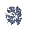 3rmaC 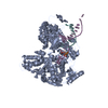 3rmcC 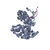 3rmdC 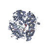 2dy4S S: Starting model for refinement C: citing same article ( |
|---|---|
| Similar structure data |


-Links
 PDBj
PDBj


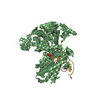
- Assembly
| Deposited unit | 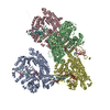
| ||||||||
|---|---|---|---|---|---|---|---|---|---|
| 1 | 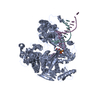
| ||||||||
| 2 | 
| ||||||||
| 3 | 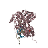
| ||||||||
| 4 | 
| ||||||||
| Unit cell |
|
-Components
| #1: Protein | Mass: 105069.586 Da / Num. of mol.: 4 / Mutation: D222A, D327A Source method: isolated from a genetically manipulated source Source: (gene. exp.) Enterobacteria phage RB69 (virus) / Gene: 43, gp43 / Production host:  #2: DNA chain | Mass: 5560.596 Da / Num. of mol.: 4 / Source method: obtained synthetically / Details: DNA template #3: DNA chain | Mass: 4271.778 Da / Num. of mol.: 4 / Source method: obtained synthetically / Details: DNA primer #4: Chemical | ChemComp-SO4 / |   Mass: 96.063 Da / Num. of mol.: 1 / Source method: obtained synthetically / Formula: SO4 Mass: 96.063 Da / Num. of mol.: 1 / Source method: obtained synthetically / Formula: SO4#5: Water | ChemComp-HOH / |  Mass: 18.015 Da / Num. of mol.: 500 / Source method: isolated from a natural source / Formula: H2O Mass: 18.015 Da / Num. of mol.: 500 / Source method: isolated from a natural source / Formula: H2O |
|---|
-Experimental details
-Experiment
| Experiment | Method: X-RAY DIFFRACTION / Number of used crystals: 1 |
|---|
- Sample preparation
| Crystal | Density Matthews: 2.92 Å3/Da / Density % sol: 57.95 % |
|---|---|
| Crystal grow | Temperature: 297 K / Method: vapor diffusion, hanging drop / pH: 7.2 Details: 6.5 % PEG 2000 MME, 100 mM Na acetate, 150 mM MgSO4, 100 mM Hepes pH 7.2, 6% glycerol and 2 mM beta-mercaptoethanol, vapor diffusion, hanging drop, temperature 297K |
-Data collection
| Diffraction | Mean temperature: 100 K | |||||||||||||||||||||||||||||||||||||||||||||||||||||||||||||||||||||||||||||
|---|---|---|---|---|---|---|---|---|---|---|---|---|---|---|---|---|---|---|---|---|---|---|---|---|---|---|---|---|---|---|---|---|---|---|---|---|---|---|---|---|---|---|---|---|---|---|---|---|---|---|---|---|---|---|---|---|---|---|---|---|---|---|---|---|---|---|---|---|---|---|---|---|---|---|---|---|---|---|
| Diffraction source | Source: SYNCHROTRON / Site: APS  / Beamline: 23-ID-D / Wavelength: 0.991873 Å / Beamline: 23-ID-D / Wavelength: 0.991873 Å | |||||||||||||||||||||||||||||||||||||||||||||||||||||||||||||||||||||||||||||
| Detector | Type: MARMOSAIC 300 mm CCD / Detector: CCD / Date: Aug 3, 2006 / Details: ADJUSTABLE FOCUSING MIRRORS IN K-B GEOMETRY | |||||||||||||||||||||||||||||||||||||||||||||||||||||||||||||||||||||||||||||
| Radiation | Monochromator: DOUBLE CRYSTAL CRYO-COOLED SI(111) / Protocol: SINGLE WAVELENGTH / Monochromatic (M) / Laue (L): M / Scattering type: x-ray | |||||||||||||||||||||||||||||||||||||||||||||||||||||||||||||||||||||||||||||
| Radiation wavelength | Wavelength: 0.991873 Å / Relative weight: 1 | |||||||||||||||||||||||||||||||||||||||||||||||||||||||||||||||||||||||||||||
| Reflection | Resolution: 2.65→50 Å / Num. obs: 151989 / % possible obs: 98.4 % / Observed criterion σ(I): 0 / Redundancy: 3.9 % / Rmerge(I) obs: 0.076 / Χ2: 1.043 / Net I/σ(I): 15.5 | |||||||||||||||||||||||||||||||||||||||||||||||||||||||||||||||||||||||||||||
| Reflection shell |
|
- Processing
| Software |
| ||||||||||||||||||||||||||||||||||||
|---|---|---|---|---|---|---|---|---|---|---|---|---|---|---|---|---|---|---|---|---|---|---|---|---|---|---|---|---|---|---|---|---|---|---|---|---|---|
| Refinement | Starting model: pdb entry 2DY4 Resolution: 2.65→50 Å / Occupancy max: 1 / Occupancy min: 0 / σ(F): 1 / Stereochemistry target values: Engh & Huber
| ||||||||||||||||||||||||||||||||||||
| Solvent computation | Bsol: 55.8807 Å2 | ||||||||||||||||||||||||||||||||||||
| Displacement parameters | Biso max: 172.06 Å2 / Biso mean: 70.1773 Å2 / Biso min: 7.77 Å2
| ||||||||||||||||||||||||||||||||||||
| Refinement step | Cycle: LAST / Resolution: 2.65→50 Å
| ||||||||||||||||||||||||||||||||||||
| Refine LS restraints |
| ||||||||||||||||||||||||||||||||||||
| Xplor file |
|
