 Movie
Movie Controller

[English] 日本語
 Yorodumi
- PDB-3rid: X-ray structure of the C-terminal swapped dimer of P114A variant ... -
+ Open data

- Basic information
| Entry | Database: PDB / ID: 3rid | ||||||
|---|---|---|---|---|---|---|---|
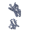
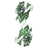
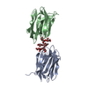
| Title | X-ray structure of the C-terminal swapped dimer of P114A variant of Ribonuclease A | ||||||
 Components Components | Ribonuclease pancreatic | ||||||
 Keywords Keywords | HYDROLASE / ribonuclease fold | ||||||
| Function / homology |  Function and homology information Function and homology informationpancreatic ribonuclease / ribonuclease A activity / RNA nuclease activity / nucleic acid binding / defense response to Gram-positive bacterium / hydrolase activity / extracellular region Similarity search - Function | ||||||
| Biological species |  | ||||||
| Method |  X-RAY DIFFRACTION / MOLECULAR REPLACEMENT / Resolution: 2.18 Å X-RAY DIFFRACTION / MOLECULAR REPLACEMENT / Resolution: 2.18 Å | ||||||
 Authors Authors | Merlino, A. / Balsamo, A. / Mazzarella, L. / Sica, F. | ||||||
 Citation Citation | Journal: Biochimie / Year: 2012 Title: Chain termini cross-talk in the swapping process of bovine pancreatic ribonuclease. Authors: Merlino, A. / Picone, D. / Ercole, C. / Balsamo, A. / Sica, F. | ||||||
| History |
|
- Structure visualization
| Structure viewer | Molecule: MolmilJmol/JSmol |
|---|
- Downloads & links
-Download
| PDBx/mmCIF format | 3rid.cif.gz | 122.6 KB | Display | PDBx/mmCIF format |
|---|---|---|---|---|
| PDB format | pdb3rid.ent.gz | 96.6 KB | Display | PDB format |
| PDBx/mmJSON format | 3rid.json.gz | Tree view | PDBx/mmJSON format | |
| Others |  Other downloads Other downloads |
-Validation report
| Arichive directory | https://data.pdbj.org/pub/pdb/validation_reports/ri/3ridftp://data.pdbj.org/pub/pdb/validation_reports/ri/3rid | HTTPS FTP |
|---|
-Related structure data
| Related structure data | 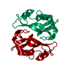 3rh1C 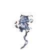 1f0vS S: Starting model for refinement C: citing same article ( |
|---|---|
| Similar structure data |


-Links
 PDBj
PDBj

- Assembly
| Deposited unit | 
| ||||||||
|---|---|---|---|---|---|---|---|---|---|
| 1 | 
| ||||||||
| 2 | 
| ||||||||
| Unit cell |
|
-Components
| #1: Protein | Mass: 13682.289 Da / Num. of mol.: 4 / Mutation: P114A Source method: isolated from a genetically manipulated source Source: (gene. exp.)  #2: Chemical | ChemComp-CGP /   Mass: 556.423 Da / Num. of mol.: 4 / Source method: obtained synthetically / Formula: C19H25N8O10P Mass: 556.423 Da / Num. of mol.: 4 / Source method: obtained synthetically / Formula: C19H25N8O10P#3: Chemical | ChemComp-PO4 /   Mass: 94.971 Da / Num. of mol.: 4 / Source method: obtained synthetically / Formula: PO4 Mass: 94.971 Da / Num. of mol.: 4 / Source method: obtained synthetically / Formula: PO4#4: Water | ChemComp-HOH / |  Mass: 18.015 Da / Num. of mol.: 546 / Source method: isolated from a natural source / Formula: H2O Mass: 18.015 Da / Num. of mol.: 546 / Source method: isolated from a natural source / Formula: H2OHas protein modification | Y | |
|---|
-Experimental details
-Experiment
| Experiment | Method: X-RAY DIFFRACTION / Number of used crystals: 1 |
|---|
- Sample preparation
| Crystal | Density Matthews: 3.41 Å3/Da / Density % sol: 63.98 % |
|---|---|
| Crystal grow | Temperature: 293 K / Method: vapor diffusion, sitting drop / pH: 6.5 Details: 17-19% PEG 20000, 0.1M cacodylate, 100-150 mg/ml trehalose, 11mM dCpdG, pH 6.5, VAPOR DIFFUSION, SITTING DROP, temperature 293.0K |
-Data collection
| Diffraction | Mean temperature: 100 K |
|---|---|
| Diffraction source | Source: ROTATING ANODE / Type: ENRAF-NONIUS FR571 / Wavelength: 1.5418 Å |
| Detector | Type: RIGAKU SATURN 944+ / Detector: CCD / Date: Nov 27, 2007 / Details: mirrors |
| Radiation | Monochromator: GRAPHITE / Protocol: SINGLE WAVELENGTH / Monochromatic (M) / Laue (L): M / Scattering type: x-ray |
| Radiation wavelength | Wavelength: 1.5418 Å / Relative weight: 1 |
| Reflection | Resolution: 2.18→30 Å / Num. all: 38571 / Num. obs: 38571 / % possible obs: 99.6 % / Observed criterion σ(F): 0 / Observed criterion σ(I): 0 / Rmerge(I) obs: 0.074 / Net I/σ(I): 14.8 |
| Reflection shell | Resolution: 2.18→2.26 Å / Rmerge(I) obs: 0.313 / Mean I/σ(I) obs: 4.3 / % possible all: 99.4 |
- Processing
| Software |
| ||||||||||||||||||||
|---|---|---|---|---|---|---|---|---|---|---|---|---|---|---|---|---|---|---|---|---|---|
| Refinement | Method to determine structure: MOLECULAR REPLACEMENT Starting model: PDB code 1F0V Resolution: 2.18→20 Å / Cross valid method: THROUGHOUT / σ(F): 2 / Stereochemistry target values: Engh & Huber
| ||||||||||||||||||||
| Refinement step | Cycle: LAST / Resolution: 2.18→20 Å
|
