 Movie
Movie Controller

[English] 日本語
 Yorodumi
- PDB-3rfx: Crystal structure of uronate dehydrogenase from Agrobacterium tum... -
+ Open data

- Basic information
| Entry | Database: PDB / ID: 3rfx | ||||||
|---|---|---|---|---|---|---|---|
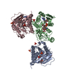
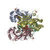
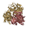
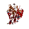
| Title | Crystal structure of uronate dehydrogenase from Agrobacterium tumefaciens, Y136A mutant complexed with NAD | ||||||
 Components Components | Uronate dehydrogenase | ||||||
 Keywords Keywords | OXIDOREDUCTASE / active site mutant / Rossmann fold / NAD | ||||||
| Function / homology |  Function and homology information Function and homology informationuronate dehydrogenase / uronate dehydrogenase activity / nucleotide binding Similarity search - Function | ||||||
| Biological species |  Agrobacterium tumefaciens (bacteria) Agrobacterium tumefaciens (bacteria) | ||||||
| Method |  X-RAY DIFFRACTION / SYNCHROTRON / MOLECULAR REPLACEMENT / Resolution: 1.9 Å X-RAY DIFFRACTION / SYNCHROTRON / MOLECULAR REPLACEMENT / Resolution: 1.9 Å | ||||||
 Authors Authors | Parkkinen, T. / Rouvinen, J. | ||||||
 Citation Citation | Journal: J.Biol.Chem. / Year: 2011 Title: Crystal Structure of Uronate Dehydrogenase from Agrobacterium tumefaciens. Authors: Parkkinen, T. / Boer, H. / Janis, J. / Andberg, M. / Penttila, M. / Koivula, A. / Rouvinen, J. | ||||||
| History |
|
- Structure visualization
| Structure viewer | Molecule: MolmilJmol/JSmol |
|---|
- Downloads & links
-Download
| PDBx/mmCIF format | 3rfx.cif.gz | 184.5 KB | Display | PDBx/mmCIF format |
|---|---|---|---|---|
| PDB format | pdb3rfx.ent.gz | 146.6 KB | Display | PDB format |
| PDBx/mmJSON format | 3rfx.json.gz | Tree view | PDBx/mmJSON format | |
| Others |  Other downloads Other downloads |
-Validation report
| Arichive directory | https://data.pdbj.org/pub/pdb/validation_reports/rf/3rfxftp://data.pdbj.org/pub/pdb/validation_reports/rf/3rfx | HTTPS FTP |
|---|
-Related structure data
| Related structure data |  3rftSC  3rfvC S: Starting model for refinement C: citing same article ( |
|---|---|
| Similar structure data |


-Links
 PDBj
PDBj- Assembly
| Deposited unit | 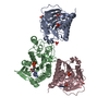
| |||||||||
|---|---|---|---|---|---|---|---|---|---|---|
| 1 | 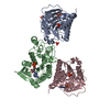
| |||||||||
| Unit cell |
| |||||||||
| Components on special symmetry positions |
|
-Components
| #1: Protein | Mass: 29186.078 Da / Num. of mol.: 3 / Mutation: Y136A Source method: isolated from a genetically manipulated source Source: (gene. exp.) Agrobacterium tumefaciens (bacteria) / Strain: C58 / ATCC 33970 / Gene: udh, AGR_L_3333, Atu3143 / Production host:  #2: Chemical |   Mass: 663.425 Da / Num. of mol.: 3 / Source method: obtained synthetically / Formula: C21H27N7O14P2 / Comment: NAD*YM Mass: 663.425 Da / Num. of mol.: 3 / Source method: obtained synthetically / Formula: C21H27N7O14P2 / Comment: NAD*YM#3: Chemical | ChemComp-PO4 /   Mass: 94.971 Da / Num. of mol.: 6 / Source method: obtained synthetically / Formula: PO4 Mass: 94.971 Da / Num. of mol.: 6 / Source method: obtained synthetically / Formula: PO4#4: Water | ChemComp-HOH / |  Mass: 18.015 Da / Num. of mol.: 896 / Source method: isolated from a natural source / Formula: H2O Mass: 18.015 Da / Num. of mol.: 896 / Source method: isolated from a natural source / Formula: H2O |
|---|
-Experimental details
-Experiment
| Experiment | Method: X-RAY DIFFRACTION / Number of used crystals: 1 |
|---|
- Sample preparation
| Crystal | Density Matthews: 3.93 Å3/Da / Density % sol: 68.72 % |
|---|---|
| Crystal grow | Temperature: 298 K / Method: vapor diffusion, hanging drop / pH: 8.5 Details: 1.8 M ammonium phosphate, Tris-HCl, pH 8.5, VAPOR DIFFUSION, HANGING DROP, temperature 298K |
-Data collection
| Diffraction | Mean temperature: 100 K |
|---|---|
| Diffraction source | Source: SYNCHROTRON / Site: ESRF  / Beamline: ID23-1 / Wavelength: 0.97 / Beamline: ID23-1 / Wavelength: 0.97 |
| Detector | Type: ADSC QUANTUM 315r / Detector: CCD / Date: Jan 1, 2010 |
| Radiation | Protocol: SINGLE WAVELENGTH / Monochromatic (M) / Laue (L): M / Scattering type: x-ray |
| Radiation wavelength | Wavelength: 0.97 Å / Relative weight: 1 |
| Reflection | Resolution: 1.9→50 Å / Num. all: 110282 / Num. obs: 107609 / % possible obs: 97.6 % |
| Reflection shell | Resolution: 1.9→1.95 Å / Redundancy: 11 % / Rmerge(I) obs: 0.382 / Mean I/σ(I) obs: 6.1 / Num. unique all: 7980 / % possible all: 98.9 |
- Processing
| Software |
| |||||||||||||||||||||||||||||||||||||||||||||||||||||||||||||||||||||||||||||
|---|---|---|---|---|---|---|---|---|---|---|---|---|---|---|---|---|---|---|---|---|---|---|---|---|---|---|---|---|---|---|---|---|---|---|---|---|---|---|---|---|---|---|---|---|---|---|---|---|---|---|---|---|---|---|---|---|---|---|---|---|---|---|---|---|---|---|---|---|---|---|---|---|---|---|---|---|---|---|
| Refinement | Method to determine structure: MOLECULAR REPLACEMENT Starting model: PDB ENTRY 3RFT Resolution: 1.9→47.515 Å / SU ML: 0.17 / σ(F): 1.99 / Phase error: 16.36
| |||||||||||||||||||||||||||||||||||||||||||||||||||||||||||||||||||||||||||||
| Solvent computation | Shrinkage radii: 0.9 Å / VDW probe radii: 1.11 Å / Solvent model: FLAT BULK SOLVENT MODEL / Bsol: 36.928 Å2 / ksol: 0.364 e/Å3 | |||||||||||||||||||||||||||||||||||||||||||||||||||||||||||||||||||||||||||||
| Displacement parameters |
| |||||||||||||||||||||||||||||||||||||||||||||||||||||||||||||||||||||||||||||
| Refinement step | Cycle: LAST / Resolution: 1.9→47.515 Å
| |||||||||||||||||||||||||||||||||||||||||||||||||||||||||||||||||||||||||||||
| Refine LS restraints |
| |||||||||||||||||||||||||||||||||||||||||||||||||||||||||||||||||||||||||||||
| LS refinement shell |
|
