 Movie
Movie Controller

[English] 日本語
 Yorodumi
- PDB-3rd6: Crystal structure of Mll3558 protein from Rhizobium loti. Northea... -
+ Open data

- Basic information
| Entry | Database: PDB / ID: 3rd6 | ||||||
|---|---|---|---|---|---|---|---|
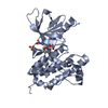
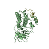
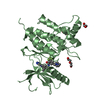
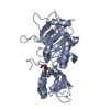
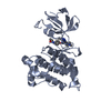
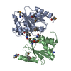
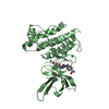
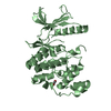
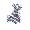
| Title | Crystal structure of Mll3558 protein from Rhizobium loti. Northeast Structural Genomics Consortium target id MlR403 | ||||||
 Components Components | Mll3558 protein | ||||||
 Keywords Keywords | Structural Genomics / Unknown function / PSI-Biology / Northeast Structural Genomics Consortium / NESG | ||||||
| Function / homology | Activator of Hsp90 ATPase homologue 1-like / Activator of Hsp90 ATPase homolog 1-like protein / START domain / Alpha-D-Glucose-1,6-Bisphosphate; Chain A, domain 4 / START-like domain superfamily / 2-Layer Sandwich / Alpha Beta / Mll3558 protein Function and homology information Function and homology information | ||||||
| Biological species |  Mesorhizobium loti (bacteria) Mesorhizobium loti (bacteria) | ||||||
| Method |  X-RAY DIFFRACTION / SYNCHROTRON / SAD / Resolution: 2.8 Å X-RAY DIFFRACTION / SYNCHROTRON / SAD / Resolution: 2.8 Å | ||||||
 Authors Authors | Seetharaman, J. / Chen, Y. / Wang, D. / Ciccosanti, C. / Sahdev, S. / Rost, B. / Acton, T.B. / Xiao, R. / Everett, J.K. / Montelione, G.T. ...Seetharaman, J. / Chen, Y. / Wang, D. / Ciccosanti, C. / Sahdev, S. / Rost, B. / Acton, T.B. / Xiao, R. / Everett, J.K. / Montelione, G.T. / Tong, L. / Hunt, J.F. / Northeast Structural Genomics Consortium (NESG) | ||||||
 Citation Citation | Journal: To be Published Title: Crystal structure of Mll3558 protein from Rhizobium loti. Northeast Structural Genomics Consortium target id MlR403 Authors: Seetharaman, J. / Chen, Y. / Wang, D. / Ciccosanti, C. / Sahdev, S. / Rost, B. / Acton, T.B. / Xiao, R. / Everett, J.K. / Montelione, G.T. / Tong, L. / Hunt, J.F. / Northeast Structural ...Authors: Seetharaman, J. / Chen, Y. / Wang, D. / Ciccosanti, C. / Sahdev, S. / Rost, B. / Acton, T.B. / Xiao, R. / Everett, J.K. / Montelione, G.T. / Tong, L. / Hunt, J.F. / Northeast Structural Genomics Consortium (NESG) | ||||||
| History |
|
- Structure visualization
| Structure viewer | Molecule: MolmilJmol/JSmol |
|---|
- Downloads & links
-Download
| PDBx/mmCIF format | 3rd6.cif.gz | 69.4 KB | Display | PDBx/mmCIF format |
|---|---|---|---|---|
| PDB format | pdb3rd6.ent.gz | 51.2 KB | Display | PDB format |
| PDBx/mmJSON format | 3rd6.json.gz | Tree view | PDBx/mmJSON format | |
| Others |  Other downloads Other downloads |
-Validation report
| Arichive directory | https://data.pdbj.org/pub/pdb/validation_reports/rd/3rd6ftp://data.pdbj.org/pub/pdb/validation_reports/rd/3rd6 | HTTPS FTP |
|---|
-Related structure data
| Similar structure data | |
|---|---|
| Other databases |

-Links
 PDBj
PDBj- Assembly
| Deposited unit | 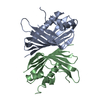
| ||||||||
|---|---|---|---|---|---|---|---|---|---|
| 1 |
| ||||||||
| 2 | 
| ||||||||
| Unit cell |
|
-Components
| #1: Protein | Mass: 18109.049 Da / Num. of mol.: 2 Source method: isolated from a genetically manipulated source Source: (gene. exp.) Mesorhizobium loti (bacteria) / Gene: mll3558 / Production host: #2: Water | ChemComp-HOH / |  Mass: 18.015 Da / Num. of mol.: 167 / Source method: isolated from a natural source / Formula: H2O Mass: 18.015 Da / Num. of mol.: 167 / Source method: isolated from a natural source / Formula: H2O |
|---|
-Experimental details
-Experiment
| Experiment | Method: X-RAY DIFFRACTION / Number of used crystals: 1 |
|---|
- Sample preparation
| Crystal | Density Matthews: 2.27 Å3/Da / Density % sol: 45.7 % |
|---|---|
| Crystal grow | Temperature: 277 K / Method: vapor diffusion, sitting drop / pH: 7 Details: 0.1M HEPES (pH 7.5), 20% PEG4000, Na Citrate, VAPOR DIFFUSION, SITTING DROP, temperature 277K |
-Data collection
| Diffraction | Mean temperature: 100 K |
|---|---|
| Diffraction source | Source: SYNCHROTRON / Site: NSLS  / Beamline: X4A / Wavelength: 0.979 Å / Beamline: X4A / Wavelength: 0.979 Å |
| Detector | Type: ADSC QUANTUM 4r / Detector: CCD / Date: Mar 7, 2011 |
| Radiation | Protocol: SINGLE WAVELENGTH / Monochromatic (M) / Laue (L): M / Scattering type: x-ray |
| Radiation wavelength | Wavelength: 0.979 Å / Relative weight: 1 |
| Reflection | Resolution: 2.8→50 Å / Num. obs: 35914 / % possible obs: 97.5 % / Observed criterion σ(F): 0 / Observed criterion σ(I): 0 / Biso Wilson estimate: 38.5 Å2 / Rmerge(I) obs: 0.051 |
| Reflection shell | Resolution: 2.8→2.86 Å / Num. unique all: 3274 / % possible all: 98 |
- Processing
| Software |
| ||||||||||||||||||||
|---|---|---|---|---|---|---|---|---|---|---|---|---|---|---|---|---|---|---|---|---|---|
| Refinement | Method to determine structure: SAD / Resolution: 2.8→25.51 Å / Rfactor Rfree error: 0.012 / Data cutoff high absF: 127326.05 / Data cutoff low absF: 0 / Isotropic thermal model: RESTRAINED / Cross valid method: THROUGHOUT / σ(F): 2 / Stereochemistry target values: Engh & Huber / Details: BULK SOLVENT MODEL USED
| ||||||||||||||||||||
| Solvent computation | Solvent model: FLAT MODEL / Bsol: 42.6943 Å2 / ksol: 0.35 e/Å3 | ||||||||||||||||||||
| Displacement parameters | Biso mean: 49.2 Å2
| ||||||||||||||||||||
| Refine analyze |
| ||||||||||||||||||||
| Refinement step | Cycle: LAST / Resolution: 2.8→25.51 Å
| ||||||||||||||||||||
| Refine LS restraints |
| ||||||||||||||||||||
| Refine LS restraints NCS | NCS model details: NONE | ||||||||||||||||||||
| LS refinement shell | Resolution: 2.8→2.98 Å / Rfactor Rfree error: 0.037 / Total num. of bins used: 6
| ||||||||||||||||||||
| Xplor file |
|
