 Movie
Movie Controller

[English] 日本語
 Yorodumi
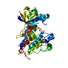
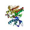
- PDB-3rc9: Crystal Structure of the K102A mutant of KijD10, a 3-ketoreductas... -
+ Open data

- Basic information
| Entry | Database: PDB / ID: 3rc9 | ||||||
|---|---|---|---|---|---|---|---|
| Title | Crystal Structure of the K102A mutant of KijD10, a 3-ketoreductase from Actinomadura kijaniata in complex with TDP-benzene and NADP | ||||||
 Components Components | Sugar 3-ketoreductase | ||||||
 Keywords Keywords | SUGAR BINDING PROTEIN / sugar biosynthesis / ketoreductase / TDP binding / NADP binding | ||||||
| Function / homology |  Function and homology information Function and homology informationdTDP-3,4-didehydro-2,6-dideoxy-alpha-D-glucose 3-reductase / antibiotic biosynthetic process / oxidoreductase activity / nucleotide binding Similarity search - Function | ||||||
| Biological species |  Actinomadura kijaniata (bacteria) Actinomadura kijaniata (bacteria) | ||||||
| Method |  X-RAY DIFFRACTION / MOLECULAR REPLACEMENT / Resolution: 1.91 Å X-RAY DIFFRACTION / MOLECULAR REPLACEMENT / Resolution: 1.91 Å | ||||||
 Authors Authors | Holden, H.M. / Kubiak, R.L. | ||||||
 Citation Citation | Journal: Biochemistry / Year: 2011 Title: Combined Structural and Functional Investigation of a C-3''-Ketoreductase Involved in the Biosynthesis of dTDP-l-Digitoxose. Authors: Kubiak, R.L. / Holden, H.M. | ||||||
| History |
|
- Structure visualization
| Structure viewer | Molecule: MolmilJmol/JSmol |
|---|
- Downloads & links
-Download
| PDBx/mmCIF format | 3rc9.cif.gz | 86.5 KB | Display | PDBx/mmCIF format |
|---|---|---|---|---|
| PDB format | pdb3rc9.ent.gz | 62.1 KB | Display | PDB format |
| PDBx/mmJSON format | 3rc9.json.gz | Tree view | PDBx/mmJSON format | |
| Others |  Other downloads Other downloads |
-Validation report
| Arichive directory | https://data.pdbj.org/pub/pdb/validation_reports/rc/3rc9ftp://data.pdbj.org/pub/pdb/validation_reports/rc/3rc9 | HTTPS FTP |
|---|
-Related structure data
| Related structure data |  3rbvSC 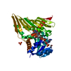 3rc1C 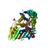 3rc2C 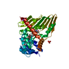 3rc7C 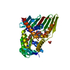 3rcbC C: citing same article ( S: Starting model for refinement |
|---|---|
| Similar structure data |


-Links
 PDBj
PDBj- Assembly
| Deposited unit | 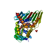
| ||||||||
|---|---|---|---|---|---|---|---|---|---|
| 1 |
| ||||||||
| Unit cell |
|
-Components
-Protein , 1 types, 1 molecules A
| #1: Protein | Mass: 39204.195 Da / Num. of mol.: 1 / Mutation: K102A Source method: isolated from a genetically manipulated source Source: (gene. exp.) Actinomadura kijaniata (bacteria) / Gene: KijD10 / Plasmid: pET28 / Production host: |
|---|
-Non-polymers , 6 types, 205 molecules 


| #2: Chemical | ChemComp-NAP /  Mass: 743.405 Da / Num. of mol.: 1 / Source method: obtained synthetically / Formula: C21H28N7O17P3 Mass: 743.405 Da / Num. of mol.: 1 / Source method: obtained synthetically / Formula: C21H28N7O17P3 | ||
|---|---|---|---|
| #3: Chemical | ChemComp-CL /  Mass: 35.453 Da / Num. of mol.: 1 / Source method: obtained synthetically / Formula: Cl Mass: 35.453 Da / Num. of mol.: 1 / Source method: obtained synthetically / Formula: Cl | ||
| #4: Chemical | ChemComp-NA /  Mass: 22.990 Da / Num. of mol.: 1 / Source method: obtained synthetically / Formula: Na Mass: 22.990 Da / Num. of mol.: 1 / Source method: obtained synthetically / Formula: Na | ||
| #5: Chemical | ChemComp-PO4 /  Mass: 94.971 Da / Num. of mol.: 1 / Source method: obtained synthetically / Formula: PO4 Mass: 94.971 Da / Num. of mol.: 1 / Source method: obtained synthetically / Formula: PO4 | ||
| #6: Chemical |  Mass: 478.284 Da / Num. of mol.: 2 / Source method: obtained synthetically / Formula: C16H20N2O11P2 Mass: 478.284 Da / Num. of mol.: 2 / Source method: obtained synthetically / Formula: C16H20N2O11P2#7: Water | ChemComp-HOH / | Mass: 18.015 Da / Num. of mol.: 199 / Source method: isolated from a natural source / Formula: H2O |
-Experimental details
-Experiment
| Experiment | Method: X-RAY DIFFRACTION / Number of used crystals: 1 |
|---|
- Sample preparation
| Crystal | Density Matthews: 3.52 Å3/Da / Density % sol: 65.11 % |
|---|---|
| Crystal grow | Temperature: 298 K / Method: vapor diffusion, hanging drop / pH: 8 Details: 1.0 M sodium/potassium phosphate, 100 mM HEPPS, 5 mM NADP, 5 mM TDP-benzene, pH 8.0, VAPOR DIFFUSION, HANGING DROP, temperature 298K |
-Data collection
| Diffraction | Mean temperature: 100 K |
|---|---|
| Diffraction source | Source: ROTATING ANODE / Type: RIGAKU RU200 / Wavelength: 1.5418 Å |
| Detector | Type: Bruker Platinum 135 / Detector: CCD / Date: Jan 22, 2011 / Details: montel mirrors |
| Radiation | Monochromator: Nickel filter / Protocol: SINGLE WAVELENGTH / Monochromatic (M) / Laue (L): M / Scattering type: x-ray |
| Radiation wavelength | Wavelength: 1.5418 Å / Relative weight: 1 |
| Reflection | Resolution: 1.91→84.76 Å / Num. all: 37962 / Num. obs: 37962 / % possible obs: 91.1 % / Observed criterion σ(F): 0 / Observed criterion σ(I): 0 / Redundancy: 4.1 % / Rmerge(I) obs: 0.067 / Rsym value: 0.067 / Net I/σ(I): 12.9 |
| Reflection shell | Resolution: 1.91→2.01 Å / Redundancy: 1.4 % / Rmerge(I) obs: 0.254 / Mean I/σ(I) obs: 2.2 / Num. unique all: 4673 / Rsym value: 0.254 / % possible all: 73.1 |
- Processing
| Software |
| |||||||||||||||||||||||||||||||||||||||||||||||||||||||||||||||||
|---|---|---|---|---|---|---|---|---|---|---|---|---|---|---|---|---|---|---|---|---|---|---|---|---|---|---|---|---|---|---|---|---|---|---|---|---|---|---|---|---|---|---|---|---|---|---|---|---|---|---|---|---|---|---|---|---|---|---|---|---|---|---|---|---|---|---|
| Refinement | Method to determine structure: MOLECULAR REPLACEMENT Starting model: model generated using binary complex, pdb entry 3RBV Resolution: 1.91→72 Å / Cor.coef. Fo:Fc: 0.931 / Cor.coef. Fo:Fc free: 0.904 / SU B: 2.958 / SU ML: 0.087 / Cross valid method: THROUGHOUT / σ(F): 0 / ESU R Free: 0.133 / Stereochemistry target values: MAXIMUM LIKELIHOOD
| |||||||||||||||||||||||||||||||||||||||||||||||||||||||||||||||||
| Solvent computation | Ion probe radii: 0.8 Å / Shrinkage radii: 0.8 Å / VDW probe radii: 1.4 Å / Solvent model: MASK | |||||||||||||||||||||||||||||||||||||||||||||||||||||||||||||||||
| Displacement parameters | Biso mean: 21.192 Å2
| |||||||||||||||||||||||||||||||||||||||||||||||||||||||||||||||||
| Refinement step | Cycle: LAST / Resolution: 1.91→72 Å
| |||||||||||||||||||||||||||||||||||||||||||||||||||||||||||||||||
| Refine LS restraints |
| |||||||||||||||||||||||||||||||||||||||||||||||||||||||||||||||||
| LS refinement shell | Resolution: 1.91→1.955 Å / Total num. of bins used: 20
|
