 Movie
Movie Controller

[English] 日本語
 Yorodumi
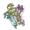
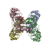
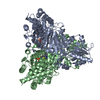
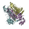
- PDB-3r5c: Pseudomonas aeruginosa DapD (PA3666) in complex with CoA and succinate -
+ Open data

- Basic information
| Entry | Database: PDB / ID: 3r5c | ||||||
|---|---|---|---|---|---|---|---|
| Title | Pseudomonas aeruginosa DapD (PA3666) in complex with CoA and succinate | ||||||
 Components Components | Tetrahydrodipicolinate N-succinyletransferase | ||||||
 Keywords Keywords | TRANSFERASE | ||||||
| Function / homology |  Function and homology information Function and homology information2,3,4,5-tetrahydropyridine-2,6-dicarboxylate N-succinyltransferase / 2,3,4,5-tetrahydropyridine-2,6-dicarboxylate N-succinyltransferase activity / : / : / magnesium ion binding / cytoplasm Similarity search - Function | ||||||
| Biological species |   Pseudomonas aeruginosa (bacteria) Pseudomonas aeruginosa (bacteria) | ||||||
| Method |  X-RAY DIFFRACTION / SYNCHROTRON / MOLECULAR REPLACEMENT / Resolution: 2.4 Å X-RAY DIFFRACTION / SYNCHROTRON / MOLECULAR REPLACEMENT / Resolution: 2.4 Å | ||||||
 Authors Authors | Sandalova, T. / Schnell, R. / Schneider, G. | ||||||
 Citation Citation | Journal: Plos One / Year: 2012 Title: Tetrahydrodipicolinate N-succinyltransferase and dihydrodipicolinate synthase from Pseudomonas aeruginosa: structure analysis and gene deletion. Authors: Schnell, R. / Oehlmann, W. / Sandalova, T. / Braun, Y. / Huck, C. / Maringer, M. / Singh, M. / Schneider, G. | ||||||
| History |
|
- Structure visualization
| Structure viewer | Molecule: MolmilJmol/JSmol |
|---|
- Downloads & links
-Download
| PDBx/mmCIF format | 3r5c.cif.gz | 198.4 KB | Display | PDBx/mmCIF format |
|---|---|---|---|---|
| PDB format | pdb3r5c.ent.gz | 159.3 KB | Display | PDB format |
| PDBx/mmJSON format | 3r5c.json.gz | Tree view | PDBx/mmJSON format | |
| Others |  Other downloads Other downloads |
-Validation report
| Arichive directory | https://data.pdbj.org/pub/pdb/validation_reports/r5/3r5cftp://data.pdbj.org/pub/pdb/validation_reports/r5/3r5c | HTTPS FTP |
|---|
-Related structure data
| Related structure data |  3qzeC  3r5aC  3r5bC  3r5dC  2rijS C: citing same article ( S: Starting model for refinement |
|---|---|
| Similar structure data |


-Links
 PDBj
PDBj


- Assembly
| Deposited unit | 
| ||||||||||||||||||||||||||||||||||||
|---|---|---|---|---|---|---|---|---|---|---|---|---|---|---|---|---|---|---|---|---|---|---|---|---|---|---|---|---|---|---|---|---|---|---|---|---|---|
| 1 |
| ||||||||||||||||||||||||||||||||||||
| Unit cell |
| ||||||||||||||||||||||||||||||||||||
| Noncrystallographic symmetry (NCS) | NCS domain:
NCS domain segments:
|
-Components
| #1: Protein | Mass: 36288.379 Da / Num. of mol.: 3 Source method: isolated from a genetically manipulated source Source: (gene. exp.) Pseudomonas aeruginosa (bacteria) / Strain: PAO1 / Gene: dapD, PA3666 / Plasmid: pET28a (Novagen) / Production host: References: UniProt: Q9Z9H2, UniProt: G3XD76*PLUS, 2,3,4,5-tetrahydropyridine-2,6-dicarboxylate N-succinyltransferase #2: Chemical |   Mass: 767.534 Da / Num. of mol.: 3 / Source method: obtained synthetically / Formula: C21H36N7O16P3S Mass: 767.534 Da / Num. of mol.: 3 / Source method: obtained synthetically / Formula: C21H36N7O16P3S#3: Chemical |   Mass: 118.088 Da / Num. of mol.: 3 / Source method: obtained synthetically / Formula: C4H6O4 Mass: 118.088 Da / Num. of mol.: 3 / Source method: obtained synthetically / Formula: C4H6O4#4: Water | ChemComp-HOH / |  Mass: 18.015 Da / Num. of mol.: 143 / Source method: isolated from a natural source / Formula: H2O Mass: 18.015 Da / Num. of mol.: 143 / Source method: isolated from a natural source / Formula: H2O |
|---|
-Experimental details
-Experiment
| Experiment | Method: X-RAY DIFFRACTION / Number of used crystals: 1 |
|---|
- Sample preparation
| Crystal | Density Matthews: 3.47 Å3/Da / Density % sol: 64.54 % |
|---|---|
| Crystal grow | Temperature: 293 K / Method: vapor diffusion, hanging drop / pH: 6.2 Details: 19-20% of PEG3350, 0.3-0.4M succinate, pH 6.2, VAPOR DIFFUSION, HANGING DROP, temperature 293K |
-Data collection
| Diffraction | Mean temperature: 100 K |
|---|---|
| Diffraction source | Source: SYNCHROTRON / Site: ESRF  / Beamline: ID14-1 / Wavelength: 0.9334 Å / Beamline: ID14-1 / Wavelength: 0.9334 Å |
| Detector | Type: ADSC QUANTUM 4r / Detector: CCD / Date: Apr 5, 2009 |
| Radiation | Monochromator: GRAPHITE / Protocol: SINGLE WAVELENGTH / Monochromatic (M) / Laue (L): M / Scattering type: x-ray |
| Radiation wavelength | Wavelength: 0.9334 Å / Relative weight: 1 |
| Reflection | Resolution: 2.4→66 Å / Num. all: 60616 / Num. obs: 60616 / % possible obs: 100 % / Observed criterion σ(F): 0 / Observed criterion σ(I): 0 / Redundancy: 6.6 % / Biso Wilson estimate: 42.1 Å2 / Rmerge(I) obs: 0.113 / Rsym value: 0.113 / Net I/σ(I): 17.1 |
| Reflection shell | Resolution: 2.4→2.53 Å / Redundancy: 6.7 % / Rmerge(I) obs: 0.654 / Mean I/σ(I) obs: 2.6 / Num. unique all: 8713 / Rsym value: 0.654 / % possible all: 100 |
- Processing
| Software |
| |||||||||||||||||||||||||||||||||||||||||||||||||||||||||||||||||||||||||||||||||||||
|---|---|---|---|---|---|---|---|---|---|---|---|---|---|---|---|---|---|---|---|---|---|---|---|---|---|---|---|---|---|---|---|---|---|---|---|---|---|---|---|---|---|---|---|---|---|---|---|---|---|---|---|---|---|---|---|---|---|---|---|---|---|---|---|---|---|---|---|---|---|---|---|---|---|---|---|---|---|---|---|---|---|---|---|---|---|---|
| Refinement | Method to determine structure: MOLECULAR REPLACEMENT Starting model: polyAla model of 2RIJ.pdb Resolution: 2.4→53.13 Å / Cor.coef. Fo:Fc: 0.923 / Cor.coef. Fo:Fc free: 0.889 / SU B: 6.981 / SU ML: 0.166 / Isotropic thermal model: isotropic / Cross valid method: THROUGHOUT / σ(I): 0 / ESU R Free: 0.24 / Stereochemistry target values: MAXIMUM LIKELIHOOD / Details: HYDROGENS HAVE BEEN ADDED IN THE RIDING POSITIONS
| |||||||||||||||||||||||||||||||||||||||||||||||||||||||||||||||||||||||||||||||||||||
| Solvent computation | Ion probe radii: 0.8 Å / Shrinkage radii: 0.8 Å / VDW probe radii: 1.4 Å / Solvent model: MASK | |||||||||||||||||||||||||||||||||||||||||||||||||||||||||||||||||||||||||||||||||||||
| Displacement parameters | Biso mean: 22.91 Å2
| |||||||||||||||||||||||||||||||||||||||||||||||||||||||||||||||||||||||||||||||||||||
| Refinement step | Cycle: LAST / Resolution: 2.4→53.13 Å
| |||||||||||||||||||||||||||||||||||||||||||||||||||||||||||||||||||||||||||||||||||||
| Refine LS restraints |
| |||||||||||||||||||||||||||||||||||||||||||||||||||||||||||||||||||||||||||||||||||||
| Refine LS restraints NCS | Dom-ID: 1 / Ens-ID: 1 / Refine-ID: X-RAY DIFFRACTION
| |||||||||||||||||||||||||||||||||||||||||||||||||||||||||||||||||||||||||||||||||||||
| LS refinement shell | Resolution: 2.4→2.462 Å / Total num. of bins used: 20
|
