 Movie
Movie Controller

[English] 日本語
 Yorodumi
- PDB-3r4y: Crystal structure of alpha-neoagarobiose hydrolase (ALPHA-NABH) f... -
+ Open data

- Basic information
| Entry | Database: PDB / ID: 3r4y | ||||||
|---|---|---|---|---|---|---|---|
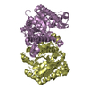
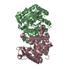
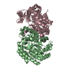
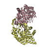
| Title | Crystal structure of alpha-neoagarobiose hydrolase (ALPHA-NABH) from Saccharophagus degradans 2-40 | ||||||
 Components Components | Glycosyl hydrolase family 32, N terminal | ||||||
 Keywords Keywords | HYDROLASE / AGAR METABOLISM / NEOAGAROBIOSE / 3 / 6-ANHYDRO-L-GALACTOSE / BIOENERGY | ||||||
| Function / homology |  Function and homology information Function and homology informationhydrolase activity, hydrolyzing O-glycosyl compounds / carbohydrate metabolic process Similarity search - Function | ||||||
| Biological species |  Saccharophagus degradans (bacteria) Saccharophagus degradans (bacteria) | ||||||
| Method |  X-RAY DIFFRACTION / SYNCHROTRON / SAD / Resolution: 2 Å X-RAY DIFFRACTION / SYNCHROTRON / SAD / Resolution: 2 Å | ||||||
 Authors Authors | Lee, S. / Lee, J.Y. / Ha, S.C. / Shin, D.H. / Kim, K.H. / Bang, W.G. / Kim, S.H. / Choi, I.G. | ||||||
 Citation Citation | Journal: Biochem.Biophys.Res.Commun. / Year: 2011 Title: Crystal structure of a key enzyme in the agarolytic pathway, alpha-neoagarobiose hydrolase from Saccharophagus degradans 2-40 Authors: Ha, S.C. / Lee, S. / Lee, J. / Kim, H.T. / Ko, H.J. / Kim, K.H. / Choi, I.G. | ||||||
| History |
|
- Structure visualization
| Structure viewer | Molecule: MolmilJmol/JSmol |
|---|
- Downloads & links
-Download
| PDBx/mmCIF format | 3r4y.cif.gz | 156.3 KB | Display | PDBx/mmCIF format |
|---|---|---|---|---|
| PDB format | pdb3r4y.ent.gz | 124.4 KB | Display | PDB format |
| PDBx/mmJSON format | 3r4y.json.gz | Tree view | PDBx/mmJSON format | |
| Others |  Other downloads Other downloads |
-Validation report
| Arichive directory | https://data.pdbj.org/pub/pdb/validation_reports/r4/3r4yftp://data.pdbj.org/pub/pdb/validation_reports/r4/3r4y | HTTPS FTP |
|---|
-Related structure data


-Links
 PDBj
PDBj- Assembly
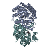
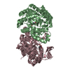
| Deposited unit | 
| ||||||||
|---|---|---|---|---|---|---|---|---|---|
| 1 |
| ||||||||
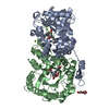
| Unit cell |
| ||||||||
| Components on special symmetry positions |
|
-Components
| #1: Protein | Mass: 42439.934 Da / Num. of mol.: 2 Source method: isolated from a genetically manipulated source Source: (gene. exp.) Saccharophagus degradans (bacteria) / Strain: 2-40 / Gene: Sde_2657 / Plasmid: PJL / Production host: References: UniProt: Q21HB2, Hydrolases; Glycosylases; Glycosidases, i.e. enzymes that hydrolyse O- and S-glycosyl compounds #2: Water | ChemComp-HOH / |  Mass: 18.015 Da / Num. of mol.: 303 / Source method: isolated from a natural source / Formula: H2O Mass: 18.015 Da / Num. of mol.: 303 / Source method: isolated from a natural source / Formula: H2O |
|---|
-Experimental details
-Experiment
| Experiment | Method: X-RAY DIFFRACTION / Number of used crystals: 1 |
|---|
- Sample preparation
| Crystal | Density Matthews: 2.6 Å3/Da / Density % sol: 52.61 % |
|---|---|
| Crystal grow | Temperature: 295 K / Method: vapor diffusion, sitting drop / pH: 7 Details: 2.4M SODIUM MALONATE, pH 7.0, VAPOR DIFFUSION, SITTING DROP, temperature 295K |
-Data collection
| Diffraction | Mean temperature: 100 K |
|---|---|
| Diffraction source | Source: SYNCHROTRON / Site: ALS  / Beamline: 5.0.2 / Wavelength: 0.9784 Å / Beamline: 5.0.2 / Wavelength: 0.9784 Å |
| Detector | Type: ADSC QUANTUM 315r / Detector: CCD / Date: Jan 20, 2009 |
| Radiation | Monochromator: DOUBLE-CRYSTAL, SI(111) / Protocol: SINGLE WAVELENGTH / Monochromatic (M) / Laue (L): M / Scattering type: x-ray |
| Radiation wavelength | Wavelength: 0.9784 Å / Relative weight: 1 |
| Reflection | Resolution: 1.98→50 Å / Num. obs: 55856 / % possible obs: 98.4 % / Observed criterion σ(I): -3 / Redundancy: 3.8 % / Rmerge(I) obs: 0.086 / Net I/σ(I): 25.3 |
| Reflection shell | Resolution: 1.98→2.05 Å / Redundancy: 3.9 % / Rmerge(I) obs: 0.46 / Mean I/σ(I) obs: 4.1 / % possible all: 98.1 |
- Processing
| Software |
| ||||||||||||||||||||||||||||||||||||||||||||||||||||||||||||||||||||||||||||||||||||||||||
|---|---|---|---|---|---|---|---|---|---|---|---|---|---|---|---|---|---|---|---|---|---|---|---|---|---|---|---|---|---|---|---|---|---|---|---|---|---|---|---|---|---|---|---|---|---|---|---|---|---|---|---|---|---|---|---|---|---|---|---|---|---|---|---|---|---|---|---|---|---|---|---|---|---|---|---|---|---|---|---|---|---|---|---|---|---|---|---|---|---|---|---|
| Refinement | Method to determine structure: SAD / Resolution: 2→19.69 Å / Cor.coef. Fo:Fc: 0.959 / Cor.coef. Fo:Fc free: 0.934 / SU B: 3.279 / SU ML: 0.094 / Cross valid method: THROUGHOUT / ESU R Free: 0.145 / Stereochemistry target values: MAXIMUM LIKELIHOOD
| ||||||||||||||||||||||||||||||||||||||||||||||||||||||||||||||||||||||||||||||||||||||||||
| Solvent computation | Ion probe radii: 0.8 Å / Shrinkage radii: 0.8 Å / VDW probe radii: 1.4 Å / Solvent model: BABINET MODEL WITH MASK | ||||||||||||||||||||||||||||||||||||||||||||||||||||||||||||||||||||||||||||||||||||||||||
| Displacement parameters | Biso mean: 23.83 Å2
| ||||||||||||||||||||||||||||||||||||||||||||||||||||||||||||||||||||||||||||||||||||||||||
| Refinement step | Cycle: LAST / Resolution: 2→19.69 Å
| ||||||||||||||||||||||||||||||||||||||||||||||||||||||||||||||||||||||||||||||||||||||||||
| Refine LS restraints |
| ||||||||||||||||||||||||||||||||||||||||||||||||||||||||||||||||||||||||||||||||||||||||||
| LS refinement shell | Resolution: 2→2.05 Å / Total num. of bins used: 20
|
