 Movie
Movie Controller


+ Open data

- Basic information
| Entry | Database: PDB / ID: 3r26 | ||||||
|---|---|---|---|---|---|---|---|
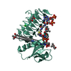
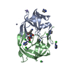
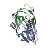
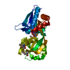
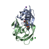
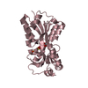
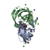
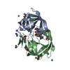
| Title | Perrhenate Binding to Molybdate Binding Protein | ||||||
 Components Components | Molybdate-binding periplasmic protein | ||||||
 Keywords Keywords | PROTEIN BINDING | ||||||
| Function / homology |  Function and homology information Function and homology informationresponse to chromate / tungstate ion transport / ABC-type molybdate transporter activity / tungstate binding / molybdate ion binding / molybdate ion transport / molybdenum ion binding / ATP-binding cassette (ABC) transporter complex, substrate-binding subunit-containing / outer membrane-bounded periplasmic space / membrane Similarity search - Function | ||||||
| Biological species |  | ||||||
| Method |  X-RAY DIFFRACTION / SYNCHROTRON / MOLECULAR REPLACEMENT / Resolution: 1.7 Å X-RAY DIFFRACTION / SYNCHROTRON / MOLECULAR REPLACEMENT / Resolution: 1.7 Å | ||||||
 Authors Authors | Aryal, B.P. / Brugarolas, P. / He, C. | ||||||
 Citation Citation | Journal: J.Biol.Inorg.Chem. / Year: 2012 Title: Binding of ReO(4) (-) with an engineered MoO (4) (2-)-binding protein: towards a new approach in radiopharmaceutical applications. Authors: Aryal, B.P. / Brugarolas, P. / He, C. | ||||||
| History |
|
- Structure visualization
| Structure viewer | Molecule: MolmilJmol/JSmol |
|---|
- Downloads & links
-Download
| PDBx/mmCIF format | 3r26.cif.gz | 104.3 KB | Display | PDBx/mmCIF format |
|---|---|---|---|---|
| PDB format | pdb3r26.ent.gz | 80.6 KB | Display | PDB format |
| PDBx/mmJSON format | 3r26.json.gz | Tree view | PDBx/mmJSON format | |
| Others |  Other downloads Other downloads |
-Validation report
| Summary document | 3r26_validation.pdf.gz | 791.8 KB | Display | wwPDB validaton report |
|---|---|---|---|---|
| Full document | 3r26_full_validation.pdf.gz | 792.6 KB | Display | |
| Data in XML | 3r26_validation.xml.gz | 11.6 KB | Display | |
| Data in CIF | 3r26_validation.cif.gz | 16.2 KB | Display | |
| Arichive directory | https://data.pdbj.org/pub/pdb/validation_reports/r2/3r26ftp://data.pdbj.org/pub/pdb/validation_reports/r2/3r26 | HTTPS FTP |
-Related structure data


-Links
 PDBj
PDBj- Assembly
| Deposited unit | 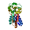
| ||||||||
|---|---|---|---|---|---|---|---|---|---|
| 1 |
| ||||||||
| Unit cell |
|
-Components
| #1: Protein | Mass: 25361.736 Da / Num. of mol.: 1 Source method: isolated from a genetically manipulated source Source: (gene. exp.) |
|---|---|
| #2: Chemical | ChemComp-REO /   Mass: 250.205 Da / Num. of mol.: 1 / Source method: obtained synthetically / Formula: O4Re Mass: 250.205 Da / Num. of mol.: 1 / Source method: obtained synthetically / Formula: O4Re |
| #3: Water | ChemComp-HOH /  Mass: 18.015 Da / Num. of mol.: 113 / Source method: isolated from a natural source / Formula: H2O Mass: 18.015 Da / Num. of mol.: 113 / Source method: isolated from a natural source / Formula: H2O |
-Experimental details
-Experiment
| Experiment | Method: X-RAY DIFFRACTION / Number of used crystals: 1 |
|---|
- Sample preparation
| Crystal | Density Matthews: 3.05 Å3/Da / Density % sol: 59.73 % |
|---|---|
| Crystal grow | Temperature: 298 K / Method: vapor diffusion, hanging drop / pH: 4.5 Details: 27.5% PEG8000, 0.1M sodium acetate, 2.5M sodium perrhenate, pH 4.5, VAPOR DIFFUSION, HANGING DROP, temperature 298K |
-Data collection
| Diffraction | Mean temperature: 77.2 K |
|---|---|
| Diffraction source | Source: SYNCHROTRON / Site: APS  / Beamline: 19-ID / Wavelength: 0.97857 Å / Beamline: 19-ID / Wavelength: 0.97857 Å |
| Detector | Type: ADSC QUANTUM 315 / Detector: CCD / Date: Feb 27, 2009 |
| Radiation | Monochromator: Si(111) / Protocol: SINGLE WAVELENGTH / Monochromatic (M) / Laue (L): M / Scattering type: x-ray |
| Radiation wavelength | Wavelength: 0.97857 Å / Relative weight: 1 |
| Reflection | Resolution: 1.7→50 Å / Num. obs: 32457 / % possible obs: 84.4 % / Redundancy: 11.1 % / Net I/σ(I): 24.3 |
| Reflection shell | Resolution: 1.7→1.79 Å / Redundancy: 10.4 % / Mean I/σ(I) obs: 6.6 / Num. unique all: 1002 / % possible all: 84.4 |
- Processing
| Software |
| ||||||||||||||||||||||||||||||||||||||||||||||||||||||||||||||||||||||||||||||||
|---|---|---|---|---|---|---|---|---|---|---|---|---|---|---|---|---|---|---|---|---|---|---|---|---|---|---|---|---|---|---|---|---|---|---|---|---|---|---|---|---|---|---|---|---|---|---|---|---|---|---|---|---|---|---|---|---|---|---|---|---|---|---|---|---|---|---|---|---|---|---|---|---|---|---|---|---|---|---|---|---|---|
| Refinement | Method to determine structure: MOLECULAR REPLACEMENT / Resolution: 1.7→50 Å / Cor.coef. Fo:Fc: 0.963 / Cor.coef. Fo:Fc free: 0.954 / SU B: 3.88 / SU ML: 0.057 / Cross valid method: THROUGHOUT / ESU R Free: 0.089 / Stereochemistry target values: MAXIMUM LIKELIHOOD Details: (1) DATA WAS COLLECTED TO THE HIGHER RESOLUTION OF 1.59 A. HOWEVER, DATA BELOW 1.7 A HAD LOW COMPLETENESS (LESS THAN 55%), AND THE SIGNAL-TO-NOISE RATIO I/SIGMA(I) WAS SIGNIFICANTLY LOW ...Details: (1) DATA WAS COLLECTED TO THE HIGHER RESOLUTION OF 1.59 A. HOWEVER, DATA BELOW 1.7 A HAD LOW COMPLETENESS (LESS THAN 55%), AND THE SIGNAL-TO-NOISE RATIO I/SIGMA(I) WAS SIGNIFICANTLY LOW (LESS THAN 1.5). THEREFORE THE DATA WAS SCALED TO 1.7 A. (2) HYDROGENS HAVE BEEN ADDED IN THE RIDING POSITIONS.
| ||||||||||||||||||||||||||||||||||||||||||||||||||||||||||||||||||||||||||||||||
| Solvent computation | Ion probe radii: 0.8 Å / Shrinkage radii: 0.8 Å / VDW probe radii: 1.4 Å / Solvent model: MASK | ||||||||||||||||||||||||||||||||||||||||||||||||||||||||||||||||||||||||||||||||
| Displacement parameters | Biso mean: 25.138 Å2
| ||||||||||||||||||||||||||||||||||||||||||||||||||||||||||||||||||||||||||||||||
| Refinement step | Cycle: LAST / Resolution: 1.7→50 Å
| ||||||||||||||||||||||||||||||||||||||||||||||||||||||||||||||||||||||||||||||||
| Refine LS restraints |
| ||||||||||||||||||||||||||||||||||||||||||||||||||||||||||||||||||||||||||||||||
| LS refinement shell | Resolution: 1.7→1.744 Å / Total num. of bins used: 20
|
