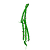
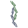
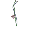
- PDB-3qwe: Crystal structure of the N-terminal domain of the GEM interacting... -
+
Open data
ID or keywords:
Loading...
-
Basic information
Entry
Database: PDB / ID: 3qwe
Title
Crystal structure of the N-terminal domain of the GEM interacting protein
Components
GEM-interacting protein
Keywords
PROTEIN BINDING / structural genomics consortium / SGC
Function / homology
Function and homology information
negative regulation of small GTPase mediated signal transduction / regulation of small GTPase mediated signal transduction / small GTPase-mediated signal transduction / CDC42 GTPase cycle / RHOA GTPase cycle / RAC1 GTPase cycle / GTPase activator activity / intracellular signal transduction / zinc ion binding / nucleoplasm ...negative regulation of small GTPase mediated signal transduction / regulation of small GTPase mediated signal transduction / small GTPase-mediated signal transduction / CDC42 GTPase cycle / RHOA GTPase cycle / RAC1 GTPase cycle / GTPase activator activity / intracellular signal transduction / zinc ion binding / nucleoplasm / plasma membrane / cytosol Similarity search - Function
Num. of mol.: 29 / Source method: obtained synthetically
-
Experimental details
-
Experiment
Experiment
Method: X-RAY DIFFRACTION / Number of used crystals: 1
-
Sample preparation
Crystal
ID
Density Matthews (Å3/Da)
Density % sol (%)
1
4
68.6
2
Crystal grow
Temperature: 293 K / Method: vapor diffusion, sitting drop / pH: 7.5 Details: 20% PEG-1500, 0.2M sodium chloride, 0.1M HEPES, 5% ethylene glycol, 3% glucose monohydrate. Crystals were de-hydrated by addition of 5% glycerol to reservoir solution, incubation over-night, ...Details: 20% PEG-1500, 0.2M sodium chloride, 0.1M HEPES, 5% ethylene glycol, 3% glucose monohydrate. Crystals were de-hydrated by addition of 5% glycerol to reservoir solution, incubation over-night, pH 7.5, vapor diffusion, sitting drop, temperature 293K, VAPOR DIFFUSION, SITTING DROP
-
Data collection
Diffraction
ID
Mean temperature (K)
Crystal-ID
1
100
1
2
100
1
Diffraction source
Source
Site
Beamline
ID
Wavelength (Å)
SYNCHROTRON
CLSI
08ID-1
1
0.92017
SYNCHROTRON
APS
19-ID
2
0.97911
Detector
Type
ID
Detector
Date
RAYONIX MX-300
1
CCD
Jan 21, 2011
ADSC QUANTUM 315
2
CCD
Dec 18, 2010
Radiation
ID
Protocol
Monochromatic (M) / Laue (L)
Scattering type
Wavelength-ID
1
SINGLEWAVELENGTH
M
x-ray
1
2
SINGLEWAVELENGTH
M
x-ray
1
Radiation wavelength
ID
Wavelength (Å)
Relative weight
1
0.92017
1
2
0.97911
1
Reflection
Resolution: 2.4→50 Å / Num. obs: 20751 / % possible obs: 99.9 % / Observed criterion σ(I): -3 / Biso Wilson estimate: 68.077 Å2 / Rmerge(I) obs: 0.065 / Net I/σ(I): 24.19
Reflection shell
Resolution (Å)
Rmerge(I) obs
Mean I/σ(I) obs
Num. measured obs
Num. unique obs
% possible all
2.4-2.46
0.945
2.2
21132
1445
99.9
2.46-2.53
0.688
3
21617
1449
100
2.53-2.6
0.57
4
21953
1409
100
2.6-2.68
0.497
4.8
22227
1354
100
2.68-2.77
0.399
6.4
22908
1353
100
2.77-2.87
0.283
9.1
23223
1306
100
2.87-2.98
0.216
12.4
22683
1229
100
2.98-3.1
0.159
16.7
23016
1212
99.9
3.1-3.24
0.122
23.2
22338
1175
100
3.24-3.39
0.106
28.2
21414
1119
100
3.39-3.58
0.088
34.3
20336
1077
100
3.58-3.79
0.068
41.8
18708
1000
100
3.79-4.06
0.062
46.7
17820
971
100
4.06-4.38
0.054
49.2
16582
904
100
4.38-4.8
0.048
52.1
15669
866
100
4.8-5.37
0.045
52.8
13989
750
100
5.37-6.2
0.047
50.9
13021
712
100
6.2-7.59
0.039
54.5
10996
611
100
7.59-10.73
0.031
57
8229
494
99.4
10.73-30
0.029
51.6
4229
315
96.6
-
Processing
Software
Name
Version
Classification
NB
XSCALE
dataprocessing
BUSTER-TNT
BUSTER2.8.0
refinement
PDB_EXTRACT
3.1
dataextraction
XDS
datareduction
DENZO
datareduction
XSCALE
datascaling
SCALEPACK
datascaling
SHELXDE
phasing
BUSTER
2.8.0
refinement
Refinement
Method to determine structure: SAD / Resolution: 2.4→30 Å / Cor.coef. Fo:Fc: 0.9215 / Cor.coef. Fo:Fc free: 0.929 / Occupancy max: 1 / Occupancy min: 1 / Cross valid method: THROUGHOUT / σ(F): 0 Details: PROGRAMS BUCCANEER, PHENIX, REFMAC, COOT AND THE MOLPROBITY SERVER WERE ALSO USED DURING REFINEMENT. FOR THIS STRUCTURE, CROSS-VALIDATION IS SUBJECT TO LARGE VARIATIONS IN RFREE, DEPENDING ...Details: PROGRAMS BUCCANEER, PHENIX, REFMAC, COOT AND THE MOLPROBITY SERVER WERE ALSO USED DURING REFINEMENT. FOR THIS STRUCTURE, CROSS-VALIDATION IS SUBJECT TO LARGE VARIATIONS IN RFREE, DEPENDING ON THE SELECTED FREE SET. THE CURRENT SET OF FREE REFLECTIONS PRODUCES AN UNREASONABLY LOW RFREE. THIS WAS CONFIRMED BY A MULTI-STEP REFINEMENT RUN THAT USED AN ALTERNATIVE SUBSET OF REFLECTIONS FOR CALCULATION OF RFREE. WE COULD NOT CONFIDENTLY INTERPRET DIFFERENCE DENSITY AT THE CURRENT MODEL'S C-TERMINUS. AMINO ACID SEQUENCE ALIGNMENT TO THE MODEL LARGELY RELIED ON THE SELENOMETHIONINE POSITIONS OBTAINED DURING PHASING. ONLY MODERATE MAP QUALITY COMBINED WITH GAPS IN THE DENSITY GIVES RISE TO SOME UNCERTAINTY IN THE AMINO ACID SEQUENCE ALIGNMENT IN SOME PARTS OF THE MODEL. DIFFRACTION IMAGES SHOW ANISOTROPIC HIGH RESOLUTION LIMITS. THE ELECTRON DENSITY MAP USED FOR MODEL BUILDING APPEARED TO BE OF SIGNIFICANTLY LOWER QUALITY THAN THE QUOTED HIGHER RESOLUTION LIMITS SUGGEST. WATER MOLECULES WERE NOT EXPLICITLY MODELED.
In the structure databanks used in Yorodumi, some data are registered as the other names, "COVID-19 virus" and "2019-nCoV". Here are the details of the virus and the list of structure data.
Jan 31, 2019. EMDB accession codes are about to change! (news from PDBe EMDB page)
EMDB accession codes are about to change! (news from PDBe EMDB page)
The allocation of 4 digits for EMDB accession codes will soon come to an end. Whilst these codes will remain in use, new EMDB accession codes will include an additional digit and will expand incrementally as the available range of codes is exhausted. The current 4-digit format prefixed with “EMD-” (i.e. EMD-XXXX) will advance to a 5-digit format (i.e. EMD-XXXXX), and so on. It is currently estimated that the 4-digit codes will be depleted around Spring 2019, at which point the 5-digit format will come into force.
The EM Navigator/Yorodumi systems omit the EMD- prefix.
Related info.:Q: What is EMD? / ID/Accession-code notation in Yorodumi/EM Navigator
Yorodumi is a browser for structure data from EMDB, PDB, SASBDB, etc.
This page is also the successor to EM Navigator detail page, and also detail information page/front-end page for Omokage search.
The word "yorodu" (or yorozu) is an old Japanese word meaning "ten thousand". "mi" (miru) is to see.
Related info.:EMDB / PDB / SASBDB / Comparison of 3 databanks / Yorodumi Search / Aug 31, 2016. New EM Navigator & Yorodumi / Yorodumi Papers / Jmol/JSmol / Function and homology information / Changes in new EM Navigator and Yorodumi
 Movie
Movie Controller

 Yorodumi
 Open data
 Basic information
 Components
 Keywords
 Function and homology information
 Homo sapiens (human)
 X-RAY DIFFRACTION /
  Authors
 Citation
 Structure visualization
 Downloads & links
 Other downloads
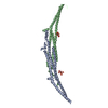
 PDBj
PDBj


 Assembly


 Num. of mol.: 29 / Source method: obtained synthetically
 Sample preparation

 Processing