 Movie
Movie Controller


+ Open data

- Basic information
| Entry | Database: PDB / ID: 3qv0 | ||||||
|---|---|---|---|---|---|---|---|
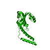
| Title | Crystal structure of Saccharomyces cerevisiae Mam33 | ||||||
 Components Components | Mitochondrial acidic protein MAM33 | ||||||
 Keywords Keywords | PROTEIN BINDING / a bowl like structure / mitochondrial oxidative phosphorylation | ||||||
| Function / homology |  Function and homology information Function and homology informationRHOC GTPase cycle / mitochondrial ribosome binding / RHOA GTPase cycle / mitochondrial ribosome assembly / aerobic respiration / cytosolic ribosome assembly / mitochondrial matrix / mitochondrion Similarity search - Function | ||||||
| Biological species |  | ||||||
| Method |  X-RAY DIFFRACTION / SYNCHROTRON / MOLECULAR REPLACEMENT / Resolution: 2.1 Å X-RAY DIFFRACTION / SYNCHROTRON / MOLECULAR REPLACEMENT / Resolution: 2.1 Å | ||||||
 Authors Authors | Jiang, Y.L. / Pu, Y.G. / Ma, X.X. / Chen, Y. / Zhou, C.Z. | ||||||
 Citation Citation | Journal: J.Struct.Biol. / Year: 2011 Title: Crystal structures and putative interface of Saccharomyces cerevisiae mitochondrial matrix proteins Mmf1 and Mam33. Authors: Pu, Y.G. / Jiang, Y.L. / Ye, X.D. / Ma, X.X. / Guo, P.C. / Lian, F.M. / Teng, Y.B. / Chen, Y. / Zhou, C.Z. | ||||||
| History |
|
- Structure visualization
| Structure viewer | Molecule: MolmilJmol/JSmol |
|---|
- Downloads & links
-Download
| PDBx/mmCIF format | 3qv0.cif.gz | 89.3 KB | Display | PDBx/mmCIF format |
|---|---|---|---|---|
| PDB format | pdb3qv0.ent.gz | 67.6 KB | Display | PDB format |
| PDBx/mmJSON format | 3qv0.json.gz | Tree view | PDBx/mmJSON format | |
| Others |  Other downloads Other downloads |
-Validation report
| Summary document | 3qv0_validation.pdf.gz | 423.6 KB | Display | wwPDB validaton report |
|---|---|---|---|---|
| Full document | 3qv0_full_validation.pdf.gz | 424.6 KB | Display | |
| Data in XML | 3qv0_validation.xml.gz | 9.2 KB | Display | |
| Data in CIF | 3qv0_validation.cif.gz | 12.2 KB | Display | |
| Arichive directory | https://data.pdbj.org/pub/pdb/validation_reports/qv/3qv0ftp://data.pdbj.org/pub/pdb/validation_reports/qv/3qv0 | HTTPS FTP |
-Related structure data
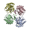
| Related structure data |  3quwC 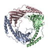 1p32S C: citing same article ( S: Starting model for refinement |
|---|---|
| Similar structure data |


-Links
 PDBj
PDBj
- Assembly
| Deposited unit | 
| ||||||||
|---|---|---|---|---|---|---|---|---|---|
| 1 | 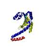
| ||||||||
| Unit cell |
|
-Components
| #1: Protein | Mass: 25875.092 Da / Num. of mol.: 1 Source method: isolated from a genetically manipulated source Source: (gene. exp.) Gene: MAM33, YIL070C / Plasmid: p28 / Production host:  |
|---|---|
| #2: Water | ChemComp-HOH /  Mass: 18.015 Da / Num. of mol.: 87 / Source method: isolated from a natural source / Formula: H2O Mass: 18.015 Da / Num. of mol.: 87 / Source method: isolated from a natural source / Formula: H2O |
-Experimental details
-Experiment
| Experiment | Method: X-RAY DIFFRACTION / Number of used crystals: 1 |
|---|
- Sample preparation
| Crystal | Density Matthews: 2.7 Å3/Da / Density % sol: 54.39 % |
|---|---|
| Crystal grow | Temperature: 289 K / Method: vapor diffusion, hanging drop / pH: 7.5 Details: 0.1 M HEPES, 20% Polyethylene glycol 4000, pH 7.5, VAPOR DIFFUSION, HANGING DROP, temperature 289K |
-Data collection
| Diffraction | Mean temperature: 100 K |
|---|---|
| Diffraction source | Source: SYNCHROTRON / Site: SSRF  / Beamline: BL17U / Wavelength: 0.9194 Å / Beamline: BL17U / Wavelength: 0.9194 Å |
| Detector | Type: RAYONIX MX-225 / Detector: CCD / Date: Mar 26, 2010 |
| Radiation | Protocol: SINGLE WAVELENGTH / Monochromatic (M) / Laue (L): M / Scattering type: x-ray |
| Radiation wavelength | Wavelength: 0.9194 Å / Relative weight: 1 |
| Reflection | Resolution: 2.1→83.97 Å / Num. obs: 16404 / % possible obs: 99.9 % / Observed criterion σ(F): -3 / Observed criterion σ(I): 0 / Redundancy: 22 % / Biso Wilson estimate: 43.8 Å2 / Rmerge(I) obs: 0.081 / Rsym value: 0.081 / Net I/σ(I): 37.7 |
| Reflection shell | Resolution: 2.1→2.18 Å / Redundancy: 21.3 % / Rmerge(I) obs: 0.393 / Mean I/σ(I) obs: 10.3 / Num. unique all: 1605 / Rsym value: 0.393 / % possible all: 100 |
- Processing
| Software |
| ||||||||||||||||||||||||||||||||||||||||||||||||||||||||||||||||||||||||||||||||||||||||||||||||||||||||||||||||||||||||||||||||||||||||||||||||||||||||||||||||||||||||||
|---|---|---|---|---|---|---|---|---|---|---|---|---|---|---|---|---|---|---|---|---|---|---|---|---|---|---|---|---|---|---|---|---|---|---|---|---|---|---|---|---|---|---|---|---|---|---|---|---|---|---|---|---|---|---|---|---|---|---|---|---|---|---|---|---|---|---|---|---|---|---|---|---|---|---|---|---|---|---|---|---|---|---|---|---|---|---|---|---|---|---|---|---|---|---|---|---|---|---|---|---|---|---|---|---|---|---|---|---|---|---|---|---|---|---|---|---|---|---|---|---|---|---|---|---|---|---|---|---|---|---|---|---|---|---|---|---|---|---|---|---|---|---|---|---|---|---|---|---|---|---|---|---|---|---|---|---|---|---|---|---|---|---|---|---|---|---|---|---|---|---|---|
| Refinement | Method to determine structure: MOLECULAR REPLACEMENT Starting model: PDB entry 1P32 Resolution: 2.1→50 Å / Cor.coef. Fo:Fc: 0.932 / Cor.coef. Fo:Fc free: 0.917 / SU B: 9.361 / SU ML: 0.117 / Cross valid method: THROUGHOUT / ESU R Free: 0.169 / Stereochemistry target values: MAXIMUM LIKELIHOOD / Details: HYDROGENS HAVE BEEN ADDED IN THE RIDING POSITIONS
| ||||||||||||||||||||||||||||||||||||||||||||||||||||||||||||||||||||||||||||||||||||||||||||||||||||||||||||||||||||||||||||||||||||||||||||||||||||||||||||||||||||||||||
| Solvent computation | Ion probe radii: 0.8 Å / Shrinkage radii: 0.8 Å / VDW probe radii: 1.4 Å / Solvent model: MASK | ||||||||||||||||||||||||||||||||||||||||||||||||||||||||||||||||||||||||||||||||||||||||||||||||||||||||||||||||||||||||||||||||||||||||||||||||||||||||||||||||||||||||||
| Displacement parameters | Biso mean: 37.453 Å2 | ||||||||||||||||||||||||||||||||||||||||||||||||||||||||||||||||||||||||||||||||||||||||||||||||||||||||||||||||||||||||||||||||||||||||||||||||||||||||||||||||||||||||||
| Refinement step | Cycle: LAST / Resolution: 2.1→50 Å
| ||||||||||||||||||||||||||||||||||||||||||||||||||||||||||||||||||||||||||||||||||||||||||||||||||||||||||||||||||||||||||||||||||||||||||||||||||||||||||||||||||||||||||
| Refine LS restraints |
| ||||||||||||||||||||||||||||||||||||||||||||||||||||||||||||||||||||||||||||||||||||||||||||||||||||||||||||||||||||||||||||||||||||||||||||||||||||||||||||||||||||||||||
| LS refinement shell | Resolution: 2.1→2.156 Å / Total num. of bins used: 20
| ||||||||||||||||||||||||||||||||||||||||||||||||||||||||||||||||||||||||||||||||||||||||||||||||||||||||||||||||||||||||||||||||||||||||||||||||||||||||||||||||||||||||||
| Refinement TLS params. | Method: refined / Refine-ID: X-RAY DIFFRACTION
| ||||||||||||||||||||||||||||||||||||||||||||||||||||||||||||||||||||||||||||||||||||||||||||||||||||||||||||||||||||||||||||||||||||||||||||||||||||||||||||||||||||||||||
| Refinement TLS group |
|
