 Movie
Movie Controller

[English] 日本語
 Yorodumi
- PDB-3qqt: Amphiphilic nanotubes in the crystal structure of a biosurfactant... -
+ Open data

- Basic information
| Entry | Database: PDB / ID: 3qqt | ||||||
|---|---|---|---|---|---|---|---|
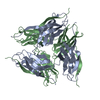
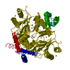
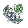
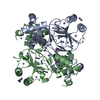
| Title | Amphiphilic nanotubes in the crystal structure of a biosurfactant protein hydrophobin HFBII | ||||||
 Components Components | Hydrophobin-2 | ||||||
 Keywords Keywords | STRUCTURAL PROTEIN / Surface active protein / amphiphile | ||||||
| Function / homology |  Function and homology information Function and homology informationconidium formation / spore wall / sporulation resulting in formation of a cellular spore / extracellular region Similarity search - Function | ||||||
| Biological species |  Trichoderma reesei (fungus) Trichoderma reesei (fungus) | ||||||
| Method |  X-RAY DIFFRACTION / SYNCHROTRON / MOLECULAR REPLACEMENT / Resolution: 1.9 Å X-RAY DIFFRACTION / SYNCHROTRON / MOLECULAR REPLACEMENT / Resolution: 1.9 Å | ||||||
 Authors Authors | Kallio, J.M. / Rouvinen, J. | ||||||
 Citation Citation | Journal: Chem.Commun.(Camb.) / Year: 2011 Title: Amphiphilic nanotubes in the crystal structure of a biosurfactant protein hydrophobin HFBII. Authors: Kallio, J.M. / Rouvinen, J. | ||||||
| History |
|
- Structure visualization
| Structure viewer | Molecule: MolmilJmol/JSmol |
|---|
- Downloads & links
-Download
| PDBx/mmCIF format | 3qqt.cif.gz | 38.9 KB | Display | PDBx/mmCIF format |
|---|---|---|---|---|
| PDB format | pdb3qqt.ent.gz | 27.7 KB | Display | PDB format |
| PDBx/mmJSON format | 3qqt.json.gz | Tree view | PDBx/mmJSON format | |
| Others |  Other downloads Other downloads |
-Validation report
| Arichive directory | https://data.pdbj.org/pub/pdb/validation_reports/qq/3qqtftp://data.pdbj.org/pub/pdb/validation_reports/qq/3qqt | HTTPS FTP |
|---|
-Related structure data
| Related structure data |  1r2mS S: Starting model for refinement |
|---|---|
| Similar structure data |


-Links
 PDBj
PDBj- Assembly
| Deposited unit | 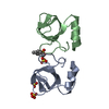
| ||||||||||||
|---|---|---|---|---|---|---|---|---|---|---|---|---|---|
| 1 | 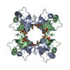
| ||||||||||||
| 2 | 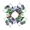
| ||||||||||||
| Unit cell |
| ||||||||||||
| Components on special symmetry positions |
|
-Components
| #1: Protein | Mass: 7201.474 Da / Num. of mol.: 2 / Source method: isolated from a natural source / Source: (natural) Trichoderma reesei (fungus) / References: UniProt: P79073#2: Chemical |   Mass: 96.063 Da / Num. of mol.: 3 / Source method: obtained synthetically / Formula: SO4 Mass: 96.063 Da / Num. of mol.: 3 / Source method: obtained synthetically / Formula: SO4#3: Chemical | ChemComp-SDS / |   Mass: 266.397 Da / Num. of mol.: 1 / Source method: obtained synthetically / Formula: C12H26O4S / Comment: detergent*YM Mass: 266.397 Da / Num. of mol.: 1 / Source method: obtained synthetically / Formula: C12H26O4S / Comment: detergent*YM#4: Water | ChemComp-HOH / |  Mass: 18.015 Da / Num. of mol.: 92 / Source method: isolated from a natural source / Formula: H2O Mass: 18.015 Da / Num. of mol.: 92 / Source method: isolated from a natural source / Formula: H2OHas protein modification | Y | |
|---|
-Experimental details
-Experiment
| Experiment | Method: X-RAY DIFFRACTION / Number of used crystals: 1 |
|---|
- Sample preparation
| Crystal | Density Matthews: 3.16 Å3/Da / Density % sol: 61.09 % |
|---|---|
| Crystal grow | Temperature: 293 K / Method: vapor diffusion, hanging drop / pH: 8.5 Details: 15% polyethylene glycol MW 2000, 0.2 M lithium sulphate, 0.1 M Tris-HCl pH 8.5, Stock solution of polystyrene nanospheres, diameter 50 nm, VAPOR DIFFUSION, HANGING DROP, temperature 293K |
-Data collection
| Diffraction | Mean temperature: 100 K | ||||||||||||||
|---|---|---|---|---|---|---|---|---|---|---|---|---|---|---|---|
| Diffraction source | Source: SYNCHROTRON / Site: EMBL/DESY, HAMBURG  / Beamline: X12 / Wavelength: 1 Å / Beamline: X12 / Wavelength: 1 Å | ||||||||||||||
| Detector | Type: MARMOSAIC 225 mm CCD / Detector: CCD / Date: Feb 15, 2007 / Details: mirrors | ||||||||||||||
| Radiation | Monochromator: Double crystal Si(111), horizontally focussing Protocol: SINGLE WAVELENGTH / Monochromatic (M) / Laue (L): M / Scattering type: x-ray | ||||||||||||||
| Radiation wavelength | Wavelength: 1 Å / Relative weight: 1 | ||||||||||||||
| Reflection | Resolution: 1.9→20 Å / Num. all: 27834 / Num. obs: 27376 / % possible obs: 98.4 % / Observed criterion σ(F): -3 / Observed criterion σ(I): -3 / Redundancy: 3.7 % / Biso Wilson estimate: 28.9 Å2 / Rmerge(I) obs: 0.089 | ||||||||||||||
| Reflection shell |
|
- Processing
| Software |
| ||||||||||||||||||||||||||||||||||||||||||||||||||||||||||||||||||||||
|---|---|---|---|---|---|---|---|---|---|---|---|---|---|---|---|---|---|---|---|---|---|---|---|---|---|---|---|---|---|---|---|---|---|---|---|---|---|---|---|---|---|---|---|---|---|---|---|---|---|---|---|---|---|---|---|---|---|---|---|---|---|---|---|---|---|---|---|---|---|---|---|
| Refinement | Method to determine structure: MOLECULAR REPLACEMENT Starting model: PDB ENTRY 1R2M Resolution: 1.9→19.22 Å / σ(F): 1.43 / Stereochemistry target values: TWIN_LSQ_F
| ||||||||||||||||||||||||||||||||||||||||||||||||||||||||||||||||||||||
| Solvent computation | Shrinkage radii: 0.9 Å / VDW probe radii: 1.11 Å / Solvent model: FLAT BULK SOLVENT MODEL / Bsol: 28.691 Å2 / ksol: 0.332 e/Å3 | ||||||||||||||||||||||||||||||||||||||||||||||||||||||||||||||||||||||
| Displacement parameters |
| ||||||||||||||||||||||||||||||||||||||||||||||||||||||||||||||||||||||
| Refinement step | Cycle: LAST / Resolution: 1.9→19.22 Å
| ||||||||||||||||||||||||||||||||||||||||||||||||||||||||||||||||||||||
| Refine LS restraints |
| ||||||||||||||||||||||||||||||||||||||||||||||||||||||||||||||||||||||
| LS refinement shell |
|
