 Movie
Movie Controller

[English] 日本語
 Yorodumi
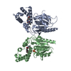
- PDB-3qp6: Crystal structure of CviR (Chromobacterium violaceum 12472) bound... -
+ Open data

- Basic information
| Entry | Database: PDB / ID: 3qp6 | ||||||
|---|---|---|---|---|---|---|---|
| Title | Crystal structure of CviR (Chromobacterium violaceum 12472) bound to C6-HSL | ||||||
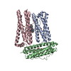
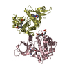
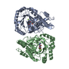
 Components Components | CviR transcriptional regulator | ||||||
 Keywords Keywords | TRANSCRIPTION / quorum sensing / agonist / antagonist / LuxR / acylated homoserine lactone / transcription factor / DNA binding protein / ligand binding domain / signal receptor / N-hexanoyl-L-homoserine lactone | ||||||
| Function / homology |  Function and homology information Function and homology information | ||||||
| Biological species |  Chromobacterium violaceum (bacteria) Chromobacterium violaceum (bacteria) | ||||||
| Method |  X-RAY DIFFRACTION / SYNCHROTRON / MOLECULAR REPLACEMENT / Resolution: 2 Å X-RAY DIFFRACTION / SYNCHROTRON / MOLECULAR REPLACEMENT / Resolution: 2 Å | ||||||
 Authors Authors | Chen, G. / Swem, L. / Swem, D. / Stauff, D. / O'Loughlin, C. / Jeffrey, P. / Bassler, B. / Hughson, F. | ||||||
 Citation Citation | Journal: Mol.Cell / Year: 2011 Title: A strategy for antagonizing quorum sensing. Authors: Chen, G. / Swem, L.R. / Swem, D.L. / Stauff, D.L. / O'Loughlin, C.T. / Jeffrey, P.D. / Bassler, B.L. / Hughson, F.M. | ||||||
| History |
|
- Structure visualization
| Structure viewer | Molecule: MolmilJmol/JSmol |
|---|
- Downloads & links
-Download
| PDBx/mmCIF format | 3qp6.cif.gz | 70 KB | Display | PDBx/mmCIF format |
|---|---|---|---|---|
| PDB format | pdb3qp6.ent.gz | 50.7 KB | Display | PDB format |
| PDBx/mmJSON format | 3qp6.json.gz | Tree view | PDBx/mmJSON format | |
| Others |  Other downloads Other downloads |
-Validation report
| Arichive directory | https://data.pdbj.org/pub/pdb/validation_reports/qp/3qp6ftp://data.pdbj.org/pub/pdb/validation_reports/qp/3qp6 | HTTPS FTP |
|---|
-Related structure data
| Related structure data | 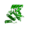 3qp1C 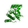 3qp2C 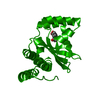 3qp4C 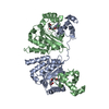 3qp5C 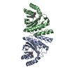 3qp8C  3qp7 C: citing same article ( S: Starting model for refinement |
|---|---|
| Similar structure data |


-Links
 PDBj
PDBj- Assembly
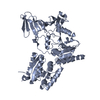
| Deposited unit | 
| |||||||||||||||
|---|---|---|---|---|---|---|---|---|---|---|---|---|---|---|---|---|
| 1 | 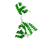
| |||||||||||||||
| Unit cell |
| |||||||||||||||
| Components on special symmetry positions |
|
-Components
| #1: Protein | Mass: 29560.760 Da / Num. of mol.: 1 Source method: isolated from a genetically manipulated source Source: (gene. exp.) Chromobacterium violaceum (bacteria) / Strain: 12472 / Gene: cviR / Plasmid: pET23b / Production host: |
|---|---|
| #2: Chemical | ChemComp-HL6 /   Mass: 199.247 Da / Num. of mol.: 1 / Source method: obtained synthetically / Formula: C10H17NO3 Mass: 199.247 Da / Num. of mol.: 1 / Source method: obtained synthetically / Formula: C10H17NO3 |
| #3: Water | ChemComp-HOH /  Mass: 18.015 Da / Num. of mol.: 241 / Source method: isolated from a natural source / Formula: H2O Mass: 18.015 Da / Num. of mol.: 241 / Source method: isolated from a natural source / Formula: H2O |
-Experimental details
-Experiment
| Experiment | Method: X-RAY DIFFRACTION / Number of used crystals: 1 |
|---|
- Sample preparation
| Crystal | Density Matthews: 2.32 Å3/Da / Density % sol: 47.04 % |
|---|---|
| Crystal grow | Temperature: 296 K / Method: vapor diffusion, hanging drop / pH: 8 Details: 100 mM imidazole, 10% w/v PEG5000 MME, pH 8.0, VAPOR DIFFUSION, HANGING DROP, temperature 296K |
-Data collection
| Diffraction | Mean temperature: 298 K |
|---|---|
| Diffraction source | Source: SYNCHROTRON / Site: NSLS  / Beamline: X25 / Wavelength: 1.0401 / Beamline: X25 / Wavelength: 1.0401 |
| Detector | Type: ADSC QUANTUM 315r / Detector: CCD / Date: Mar 6, 2008 |
| Radiation | Monochromator: Si(111) / Protocol: SINGLE WAVELENGTH / Monochromatic (M) / Laue (L): M / Scattering type: x-ray |
| Radiation wavelength | Wavelength: 1.0401 Å / Relative weight: 1 |
| Reflection | Resolution: 2→50 Å / Num. obs: 20522 / % possible obs: 99.9 % / Observed criterion σ(F): 2 / Observed criterion σ(I): 2 / Redundancy: 5.5 % / Rsym value: 0.071 / Net I/σ(I): 17.127 |
| Reflection shell | Resolution: 2→2.07 Å / Redundancy: 5.2 % / Rmerge(I) obs: 0.319 / Mean I/σ(I) obs: 4.656 / % possible all: 99.9 |
- Processing
| Software |
| ||||||||||||||||||||||||||||||||||||||||||||||||||||||||
|---|---|---|---|---|---|---|---|---|---|---|---|---|---|---|---|---|---|---|---|---|---|---|---|---|---|---|---|---|---|---|---|---|---|---|---|---|---|---|---|---|---|---|---|---|---|---|---|---|---|---|---|---|---|---|---|---|---|
| Refinement | Method to determine structure: MOLECULAR REPLACEMENT Starting model: PDB ENTRY 3QP7 3qp7 Resolution: 2→26.536 Å / SU ML: 0.23 / σ(F): 1.34 / Phase error: 20.8 / Stereochemistry target values: ML
| ||||||||||||||||||||||||||||||||||||||||||||||||||||||||
| Solvent computation | Shrinkage radii: 0.9 Å / VDW probe radii: 1.11 Å / Solvent model: FLAT BULK SOLVENT MODEL / Bsol: 27.116 Å2 / ksol: 0.323 e/Å3 | ||||||||||||||||||||||||||||||||||||||||||||||||||||||||
| Displacement parameters |
| ||||||||||||||||||||||||||||||||||||||||||||||||||||||||
| Refinement step | Cycle: LAST / Resolution: 2→26.536 Å
| ||||||||||||||||||||||||||||||||||||||||||||||||||||||||
| Refine LS restraints |
| ||||||||||||||||||||||||||||||||||||||||||||||||||||||||
| LS refinement shell |
|
