 Movie
Movie Controller
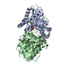

[English] 日本語
 Yorodumi
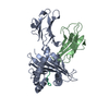
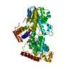
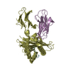
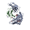
- PDB-3qmf: Crystal strucuture of an inositol monophosphatase family protein ... -
+ Open data

- Basic information
| Entry | Database: PDB / ID: 3qmf | ||||||
|---|---|---|---|---|---|---|---|
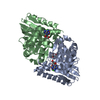
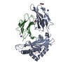
| Title | Crystal strucuture of an inositol monophosphatase family protein (SAS2203) from Staphylococcus aureus MSSA476 | ||||||
 Components Components | Inositol monophosphatase family protein | ||||||
 Keywords Keywords | HYDROLASE / FIG superfamily / IMPase fold | ||||||
| Function / homology | D-Maltodextrin-Binding Protein; domain 2 - #80 / Fructose-1,6-Bisphosphatase, subunit A, domain 1 / Fructose-1,6-Bisphosphatase; Chain A, domain 1 / D-Maltodextrin-Binding Protein; domain 2 / 2-Layer Sandwich / 3-Layer(aba) Sandwich / Alpha Beta / :  Function and homology information Function and homology information | ||||||
| Biological species |   Staphylococcus aureus (bacteria) Staphylococcus aureus (bacteria) | ||||||
| Method |  X-RAY DIFFRACTION / MOLECULAR REPLACEMENT / Resolution: 2.6 Å X-RAY DIFFRACTION / MOLECULAR REPLACEMENT / Resolution: 2.6 Å | ||||||
 Authors Authors | Bhattacharyya, S. / Dutta, D. / Ghosh, A.K. / Das, A.K. | ||||||
 Citation Citation | Journal: Biochimie / Year: 2012 Title: Crystal structure of Staphylococcal dual specific inositol monophosphatase/NADP(H) phosphatase (SAS2203) delineates the molecular basis of substrate specificity Authors: Bhattacharyya, S. / Dutta, D. / Saha, B. / Ghosh, A.K. / Das, A.K. | ||||||
| History |
|
- Structure visualization
| Structure viewer | Molecule: MolmilJmol/JSmol |
|---|
- Downloads & links
-Download
| PDBx/mmCIF format | 3qmf.cif.gz | 211.9 KB | Display | PDBx/mmCIF format |
|---|---|---|---|---|
| PDB format | pdb3qmf.ent.gz | 169.8 KB | Display | PDB format |
| PDBx/mmJSON format | 3qmf.json.gz | Tree view | PDBx/mmJSON format | |
| Others |  Other downloads Other downloads |
-Validation report
| Arichive directory | https://data.pdbj.org/pub/pdb/validation_reports/qm/3qmfftp://data.pdbj.org/pub/pdb/validation_reports/qm/3qmf | HTTPS FTP |
|---|
-Related structure data
| Related structure data |  3rydC  2p3nS C: citing same article ( S: Starting model for refinement |
|---|---|
| Similar structure data |

-Links
 PDBj
PDBj- Assembly
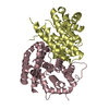
| Deposited unit | 
| ||||||||
|---|---|---|---|---|---|---|---|---|---|
| 1 |
| ||||||||
| Unit cell |
|
-Components
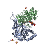
| #1: Protein | Mass: 31458.146 Da / Num. of mol.: 2 Source method: isolated from a genetically manipulated source Source: (gene. exp.) Staphylococcus aureus (bacteria) / Strain: MSSA476 / Gene: SAS2203 / Plasmid: pQE30 / Production host: #2: Chemical |   Mass: 96.063 Da / Num. of mol.: 3 / Source method: obtained synthetically / Formula: SO4 Mass: 96.063 Da / Num. of mol.: 3 / Source method: obtained synthetically / Formula: SO4#3: Water | ChemComp-HOH / |  Mass: 18.015 Da / Num. of mol.: 79 / Source method: isolated from a natural source / Formula: H2O Mass: 18.015 Da / Num. of mol.: 79 / Source method: isolated from a natural source / Formula: H2O |
|---|
-Experimental details
-Experiment
| Experiment | Method: X-RAY DIFFRACTION / Number of used crystals: 1 |
|---|
- Sample preparation
| Crystal | Density Matthews: 1.95 Å3/Da / Density % sol: 36.98 % |
|---|---|
| Crystal grow | Temperature: 298 K / Method: vapor diffusion, sitting drop / pH: 7 Details: 0.2M Lithium sulphate, 0.1M HEPES pH 7.0, 20%(w/v) PEG 3350, VAPOR DIFFUSION, SITTING DROP, temperature 298K |
-Data collection
| Diffraction | Mean temperature: 100 K |
|---|---|
| Diffraction source | Source: ROTATING ANODE / Type: RIGAKU MICROMAX-007 HF / Wavelength: 1.54 Å |
| Detector | Type: RIGAKU RAXIS IV++ / Detector: IMAGE PLATE / Date: Nov 14, 2010 / Details: mirrors |
| Radiation | Monochromator: mirror / Protocol: SINGLE WAVELENGTH / Monochromatic (M) / Laue (L): M / Scattering type: x-ray |
| Radiation wavelength | Wavelength: 1.54 Å / Relative weight: 1 |
| Reflection | Resolution: 2.597→71.894 Å / Num. all: 15862 / Num. obs: 15691 / Observed criterion σ(F): 3 / Observed criterion σ(I): 3 |
- Processing
| Software |
| |||||||||||||||||||||||||||||||||||||||||||||||||||||||||||||||||||||||||||
|---|---|---|---|---|---|---|---|---|---|---|---|---|---|---|---|---|---|---|---|---|---|---|---|---|---|---|---|---|---|---|---|---|---|---|---|---|---|---|---|---|---|---|---|---|---|---|---|---|---|---|---|---|---|---|---|---|---|---|---|---|---|---|---|---|---|---|---|---|---|---|---|---|---|---|---|---|
| Refinement | Method to determine structure: MOLECULAR REPLACEMENT Starting model: PDB ENTRY 2P3N Resolution: 2.6→19.67 Å / Cor.coef. Fo:Fc: 0.932 / Cor.coef. Fo:Fc free: 0.87 / Occupancy max: 1 / Occupancy min: 0.5 / SU B: 27.287 / SU ML: 0.27 / Cross valid method: THROUGHOUT / ESU R Free: 0.374 / Stereochemistry target values: MAXIMUM LIKELIHOOD / Details: HYDROGENS HAVE BEEN ADDED IN THE RIDING POSITIONS
| |||||||||||||||||||||||||||||||||||||||||||||||||||||||||||||||||||||||||||
| Solvent computation | Ion probe radii: 0.8 Å / Shrinkage radii: 0.8 Å / VDW probe radii: 1.4 Å / Solvent model: MASK | |||||||||||||||||||||||||||||||||||||||||||||||||||||||||||||||||||||||||||
| Displacement parameters | Biso max: 88.08 Å2 / Biso mean: 36.195 Å2 / Biso min: 2 Å2
| |||||||||||||||||||||||||||||||||||||||||||||||||||||||||||||||||||||||||||
| Refinement step | Cycle: LAST / Resolution: 2.6→19.67 Å
| |||||||||||||||||||||||||||||||||||||||||||||||||||||||||||||||||||||||||||
| Refine LS restraints |
| |||||||||||||||||||||||||||||||||||||||||||||||||||||||||||||||||||||||||||
| LS refinement shell | Resolution: 2.597→2.665 Å / Total num. of bins used: 20
| |||||||||||||||||||||||||||||||||||||||||||||||||||||||||||||||||||||||||||
| Refinement TLS params. | Method: refined / Refine-ID: X-RAY DIFFRACTION
| |||||||||||||||||||||||||||||||||||||||||||||||||||||||||||||||||||||||||||
| Refinement TLS group |
|
