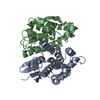
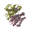
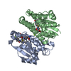
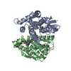
- PDB-3qkb: Crystal structure of a Protein with unknown function which belong... -
+
Open data
ID or keywords:
Loading...
-
Basic information
Entry
Database: PDB / ID: 3qkb
Title
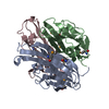
Crystal structure of a Protein with unknown function which belongs to Pfam DUF74 family (PEPE_0654) from Pediococcus pentosaceus ATCC 25745 at 2.73 A resolution
Components
Uncharacterized protein
Keywords
Structural Genomics / Unknown function / BETA/ALPHA-PROPELLER / JOINT CENTER FOR STRUCTURAL GENOMICS / JCSG / PROTEIN STRUCTURE INITIATIVE / PSI-BIOLOGY
Function / homology
UPF0145 domain superfamily / Hypothetical protein apc22750. Chain B / Translation Initiation Factor IF3 / 2-Layer Sandwich / Alpha Beta / Heavy metal-binding domain-containing protein
Function and homology information
Biological species
Pediococcus pentosaceus (bacteria)
Method
X-RAY DIFFRACTION / SYNCHROTRON / MAD / Resolution: 2.73 Å
Monochromator: SINGLE CRYSTAL SI(111) BENT MONOCHROMATOR (HORIZONTAL FOCUSING) Protocol: MAD / Monochromatic (M) / Laue (L): M / Scattering type: x-ray
Radiation wavelength
ID
Wavelength (Å)
Relative weight
1
0.91162
1
2
0.97929
1
Reflection
Resolution: 2.73→47.727 Å / Num. obs: 17422 / % possible obs: 98.8 % / Observed criterion σ(I): -3 / Biso Wilson estimate: 60.247 Å2 / Rmerge(I) obs: 0.1 / Net I/σ(I): 10.92
Reflection shell
Resolution (Å)
Rmerge(I) obs
Mean I/σ(I) obs
Num. measured obs
Num. unique obs
Diffraction-ID
% possible all
2.73-2.83
0.757
2.1
6530
1768
1
99.3
2.83-2.94
0.516
2.9
6146
1667
1
99.3
2.94-3.07
0.408
3.5
6148
1674
1
99.3
3.07-3.23
0.272
5.1
6292
1707
1
99.1
3.23-3.44
0.185
7.1
6556
1782
1
99.3
3.44-3.7
0.135
9.3
6245
1704
1
99.1
3.7-4.07
0.086
13.5
6317
1737
1
99.3
4.07-4.65
0.054
18.8
6309
1738
1
99.1
4.65-5.84
0.05
20.5
6423
1781
1
98.7
5.84-47.727
0.044
24.8
6490
1864
1
95.7
-
Phasing
Phasing
Method: MAD
-
Processing
Software
Name
Version
Classification
NB
MolProbity
3beta29
modelbuilding
PDB_EXTRACT
3.1
dataextraction
SHELX
phasing
SHARP
phasing
XSCALE
datascaling
REFMAC
5.5.0110
refinement
XDS
datareduction
SHELXD
phasing
autoSHARP
phasing
Refinement
Method to determine structure: MAD / Resolution: 2.73→47.727 Å / Cor.coef. Fo:Fc: 0.935 / Cor.coef. Fo:Fc free: 0.916 / Occupancy max: 1 / Occupancy min: 0.37 / SU B: 24.741 / SU ML: 0.238 / Cross valid method: THROUGHOUT / σ(F): 0 / ESU R: 0.782 / ESU R Free: 0.314 Stereochemistry target values: MAXIMUM LIKELIHOOD WITH PHASES Details: 1. HYDROGENS HAVE BEEN ADDED IN THE RIDING POSITIONS. 2. A MET-INHIBITION PROTOCOL WAS USED FOR SELENOMETHIONINE INCORPORATION DURING PROTEIN EXPRESSION. THE OCCUPANCY OF THE SE ATOMS IN THE ...Details: 1. HYDROGENS HAVE BEEN ADDED IN THE RIDING POSITIONS. 2. A MET-INHIBITION PROTOCOL WAS USED FOR SELENOMETHIONINE INCORPORATION DURING PROTEIN EXPRESSION. THE OCCUPANCY OF THE SE ATOMS IN THE MSE RESIDUES WAS REDUCED TO 0.75 FOR THE REDUCED SCATTERING POWER DUE TO PARTIAL S-MET INCORPORATION. 3. ATOM RECORD CONTAINS SUM OF TLS AND RESIDUAL B FACTORS. ANISOU RECORD CONTAINS SUM OF TLS AND RESIDUAL U FACTORS. 4. WATERS WERE EXCLUDED FROM AUTOMATIC TLS ASSIGNMENT.
Rfactor
Num. reflection
% reflection
Selection details
Rfree
0.2393
884
5.1 %
RANDOM
Rwork
0.2018
-
-
-
obs
0.2037
17408
98.95 %
-
Solvent computation
Ion probe radii: 0.8 Å / Shrinkage radii: 0.8 Å / VDW probe radii: 1.4 Å / Solvent model: MASK
In the structure databanks used in Yorodumi, some data are registered as the other names, "COVID-19 virus" and "2019-nCoV". Here are the details of the virus and the list of structure data.
Jan 31, 2019. EMDB accession codes are about to change! (news from PDBe EMDB page)
EMDB accession codes are about to change! (news from PDBe EMDB page)
The allocation of 4 digits for EMDB accession codes will soon come to an end. Whilst these codes will remain in use, new EMDB accession codes will include an additional digit and will expand incrementally as the available range of codes is exhausted. The current 4-digit format prefixed with “EMD-” (i.e. EMD-XXXX) will advance to a 5-digit format (i.e. EMD-XXXXX), and so on. It is currently estimated that the 4-digit codes will be depleted around Spring 2019, at which point the 5-digit format will come into force.
The EM Navigator/Yorodumi systems omit the EMD- prefix.
Related info.:Q: What is EMD? / ID/Accession-code notation in Yorodumi/EM Navigator
Yorodumi is a browser for structure data from EMDB, PDB, SASBDB, etc.
This page is also the successor to EM Navigator detail page, and also detail information page/front-end page for Omokage search.
The word "yorodu" (or yorozu) is an old Japanese word meaning "ten thousand". "mi" (miru) is to see.
Related info.:EMDB / PDB / SASBDB / Comparison of 3 databanks / Yorodumi Search / Aug 31, 2016. New EM Navigator & Yorodumi / Yorodumi Papers / Jmol/JSmol / Function and homology information / Changes in new EM Navigator and Yorodumi
 Movie
Movie Controller

 Yorodumi
 Open data
 Basic information
 Components
 Keywords
 Function and homology information
 Pediococcus pentosaceus (bacteria)
 X-RAY DIFFRACTION /
  Authors
 Citation
 Structure visualization
 Downloads & links
 Other downloads

 PDBj
PDBj Assembly
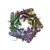
 Mass: 18.015 Da / Num. of mol.: 54 / Source method: isolated from a natural source / Formula: H2O
 Sample preparation
 / Beamline: BL11-1 / Wavelength: 0.91162,0.97929
 Processing